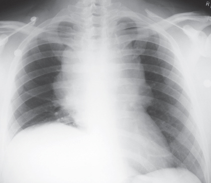
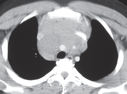
CASE 158 35-year-old man with facial swelling and flushing PA (Fig. 158.1) chest radiograph demonstrates a large, lobular, well-defined anterior mediastinal mass, which extends to both sides of the midline and is associated with elevation of the right hemidiaphragm. Contrast-enhanced chest CT (mediastinal window) (Figs. 158.2, 158.3) demonstrates a bulky anterior mediastinal mass of homogeneous soft-tissue attenuation and lobular borders. The mass encases the right brachiocephalic artery, the left common carotid artery, and the bilateral brachiocephalic veins (Fig. 158.2) and invades the superior vena cava (Fig. 158.3). Seminoma • Lymphoma • Thymoma, invasive Seminoma is an uncommon neoplasm that represents approximately 40% of primary mediastinal malignant germ cell neoplasms of a single histology. Non-seminomatous malignant germ cell neoplasms (GCNs) are aggressive neoplasms and include embryonal carcinoma, endodermal sinus (yolk sac) tumor, and choriocarcinoma. These lesions typically exhibit more than one germ cell histology and may contain foci of seminoma. Fig. 158.1 Fig. 158.2
 Clinical Presentation
 Radiologic Findings
 Diagnosis
 Differential Diagnosis
 Discussion
Background


Stay updated, free articles. Join our Telegram channel

Full access? Get Clinical Tree


