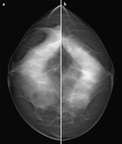
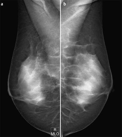
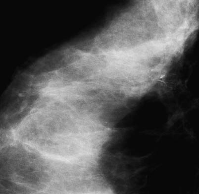
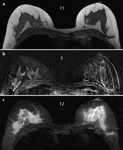
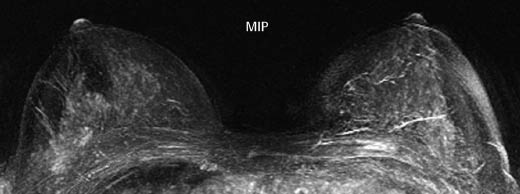
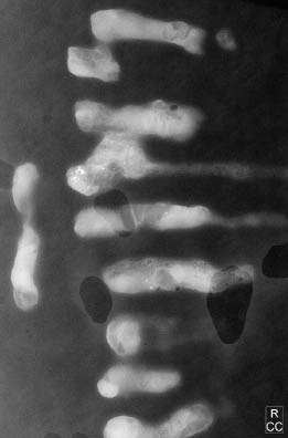
Case 16 Indication: Screening. History: No abnormalities. Risk profile: No increased risk. Age: 45 years. Fig. 16.1 a,b Digital mammography, CC view. Fig. 16.2 a,b Digital mammography, MLO view. Lumpy parenchymal texture. No resistance. No findings. Fig. 16.3 Magnification view of the right breast (CC). Fig. 16.4a -c Contrast-enhanced MR mammography. Fig. 16.5 Contrast-enhanced MR mammography. Maximum intensity projection. Please characterize mammography and MRI. What is your preliminary diagnosis? What are your next steps? This case investigated microcalcifications that were discovered in screening mammography. Ultrasound showed no abnormalities in either breast. Even with the mammography data as a guide, no echo alteration was detected in the upper outer quadrant of the right breast. Mammography demonstrated bilaterally symmetric, extremely dense parenchyma, ACR type 4. In the upper outer quadrant of the right breast a cluster of polymorphous microcalcifications was visible in a segmental orientation. Magnification mammography depicted the polymorphous character in more detail (linear and Y-shaped calcifications). No hyperdensities or architectural distortion. BI-RADS right 4/left 1. PGMI: CC view P; MLO view P. There were no pathological findings in MR mammography. In the right upper outer ROI, strong contrast uptake throughout the parenchyma meant that no focal, linear or dendritic enhancements were detected. MRI Artifact Category: right 2/left 3 MRI Density Type: 2
Clinical Findings
Ultrasound

Ultrasound
Mammography
MR Mammography
MRM score | Finding | Points |
Shape | – | 0 |
Border | – | 0 |
CM Distribution | – | 0 |
Initial Signal Intensity Increase | – | 0 |
Post-initial Signal Intensity Character | – | 0 |
MRI score (points) |
| 0 |
MRI BI-RADS |
| 1 |
 Preliminary Diagnosis
Intraductal carcinoma (occult on US and MRI), probably minimally invasive.
Clinical Findings | right 1 | left 1 |
Ultrasound | right 1 | left 1 |
Mammography | right 4 | left 1 |
MR Mammography | right 1 | left 1 |
BI-RADS Total | right 4 | left 1 |
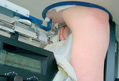
Fig. 16.6 Vacuum biopsy using a Lorad table. An atypical positioning of the arm was adopted because the calcifications were very close to the chest wall.
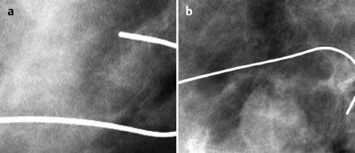
Fig. 16.7a,b Preoperative hook-wire localization, CC and MLO magnifications.
Procedure
Histopathological evaluation of the microcalcifications in the upper outer quadrant of the right breast by stereotactic vacuum biopsy (Fig. 16.6).
Histopathology of the core biopsy specimen: Invasive lobular carcinoma.
Further procedure: Preoperative hook-wire localization of the calcification cluster (Fig. 16.7a, b).
Fig. 16.8 Specimen radiography.
Histology
Invasive lobular carcinoma 12 mm in maximum diameter. Axillary lymph node status normal.
ILC* pT1 b, pN0, M0, Estrogen receptor (+), Gestagen receptor (+).
Stay updated, free articles. Join our Telegram channel

Full access? Get Clinical Tree


