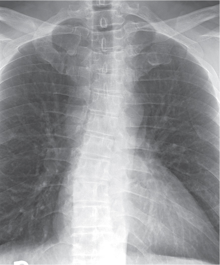
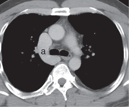
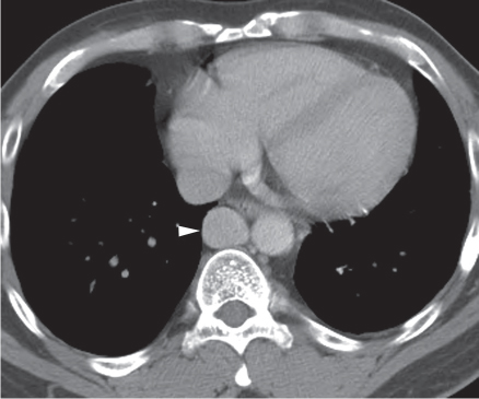
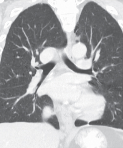
CASE 16 50-year-old man evaluated for chest and abdominal pain Coned-down PA chest radiograph (Fig. 16.1) demonstrates enlargement of the azygos arch, a right eparterial bronchus, and a left hyparterial bronchus. Axial (Figs. 16.2, 16.3) and coronal (Fig. 16.4) contrast-enhanced chest CT (mediastinal and lung windows) show enlargement of the azygos arch (a) and azygos vein (arrowhead). Note right eparterial and left hyparterial bronchi consistent with situs solitus (Fig. 16.4). Interruption of the Inferior Vena Cava with Azygos Continuation • Lymphadenopathy • Situs Ambiguous Fig. 16.1 Fig. 16.2 Fig. 16.3 Fig. 16.4
 Clinical Presentation
 Radiologic Findings
 Diagnosis
 Differential Diagnosis
 Discussion
Background
Related posts:
![]()
Stay updated, free articles. Join our Telegram channel

Full access? Get Clinical Tree


Radiology Key
Fastest Radiology Insight Engine


