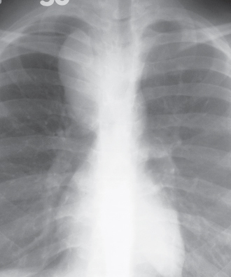
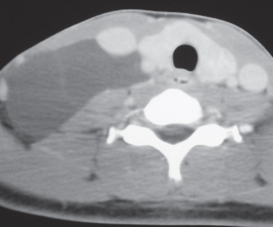
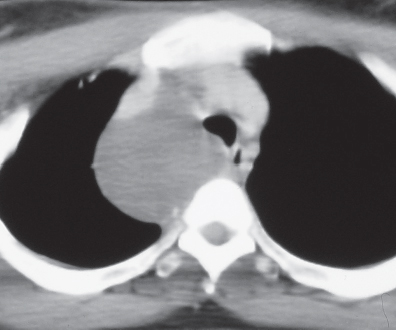
CASE 165 24-year-old woman with a palpable neck mass PA chest radiograph (Fig. 165.1) demonstrates a well-defined right middle-posterior mediastinal (paratracheal) mass that produces mass effect on the cervical and intrathoracic portions of the trachea. Contrast-enhanced chest CT (mediastinal window) (Figs. 165.2, 165.3) shows a multilocular cystic right neck mass of water attenuation contents with thin internal soft-tissue septa (Fig. 165.2) that extends into the mediastinum, insinuating between adjacent vascular structures (Fig. 165.3), and produces mass effect on the trachea and esophagus (Fig. 165.3). Lymphangioma • Congenital or Acquired Cyst • Mature Teratoma Fig. 165.1 Fig. 165.2 Fig. 165.3
 Clinical Presentation
 Radiologic Findings
 Diagnosis
 Differential Diagnosis
 Bronchogenic Cyst
 Thyroglossal Cyst
 Branchial Cleft Cyst
 Thymic Cyst
 Discussion
Background
Related posts:
![]()
Stay updated, free articles. Join our Telegram channel

Full access? Get Clinical Tree


Radiology Key
Fastest Radiology Insight Engine


