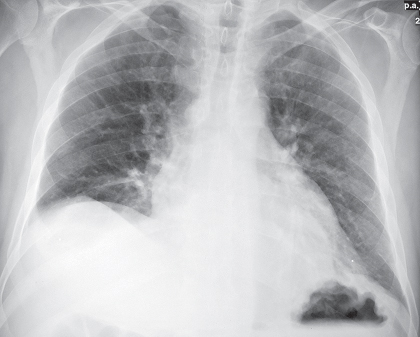
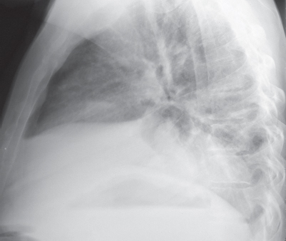
CASE 166 Asymptomatic elderly man with known cirrhosis and portal hypertension PA (Fig. 166.1) and lateral (Fig. 166.2) chest radiographs demonstrate a large retrocardiac middle/posterior mediastinal mass, basilar reticular opacities, and elevation of the right hemidiaphragm. Contrast-enhanced chest CT (mediastinal window) (Figs. 166.3, 166.4) reveals a complex paraesophageal mass composed of numerous enhancing tortuous serpiginous vascular structures. The degree of contrast enhancement within the lesion parallels that of the adjacent aorta and hemiazygos vein. Note the thickened nodular esophageal wall with minute intensely enhancing foci adjacent to the esophageal lumen and the abdominal ascites. Paraesophageal and Esophageal Varices • None Paraesophageal and esophageal varices represent dilated extrinsic and intrinsic esophageal veins, respectively. They are supplied by the left gastric vein, which arises at the portal venous confluence and courses through the gastric fundus to drain into the lower esophageal plexus veins, serving as a portosystemic collateral pathway in patients with portal hypertension. The posterior branch of the left gastric vein supplies the paraesophageal varices and the anterior branch supplies the gastroesophageal varices. Paraesophageal varices are located outside the esophageal wall, communicate with esophageal varices, and typically drain into the azygos/hemiazygos system, the subclavian/brachiocephalic system, or the inferior vena cava. Esophageal varices are located within the wall of the inferior esophagus. These collateral pathways allow decompression of the portal vein into the systemic circulation. Fig. 166.1 Fig. 166.2
 Clinical Presentation
 Radiologic Findings
 Diagnosis
 Differential Diagnosis
 Discussion
Background


Stay updated, free articles. Join our Telegram channel

Full access? Get Clinical Tree


