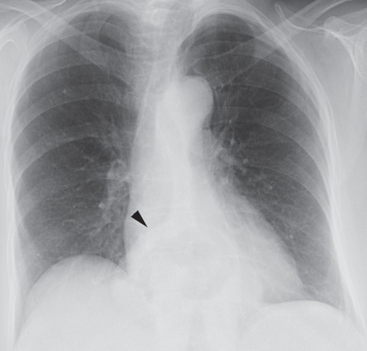
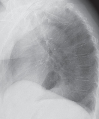
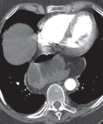
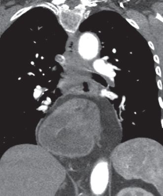
CASE 167 Middle-aged woman with recurrent epigastric pain PA (Fig. 167.1) and lateral (Fig. 167.2) chest radiographs demonstrate a moderate-size middle-posterior mediastinal (retrocardiac) mass that contains intrinsic air and fluid and produces lateral displacement of the inferior aspect of the azygoesophageal recess (arrowhead). Contrast-enhanced chest CT (mediastinal window) (Figs. 167.3, 167.4) shows a moderate hiatus hernia that contains a portion of the stomach and surrounding omental fat. Hiatus Hernia • None Hiatus hernia results from transient or permanent intrathoracic gastric herniation through an enlarged esophageal hiatus. Herniation of omentum and other portions of the gastrointestinal tract (hollow viscera and solid organs) occasionally occurs. Abdominal fluid may also herniate through the hiatus in patients with ascites. In paraesophageal hiatus hernia, the gastroesophageal junction remains in its normal location and the herniated stomach migrates above it and resides alongside the esophagus. Fig. 167.1 Fig. 167.2 Fig. 167.3 Fig. 167.4
 Clinical Presentation
 Radiologic Findings
 Diagnosis
 Differential Diagnosis
 Discussion
Background
Etiology
Related posts:
![]()
Stay updated, free articles. Join our Telegram channel

Full access? Get Clinical Tree


Radiology Key
Fastest Radiology Insight Engine


