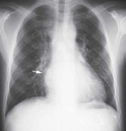
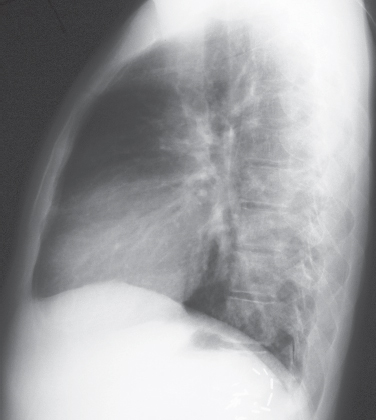
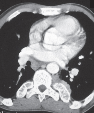
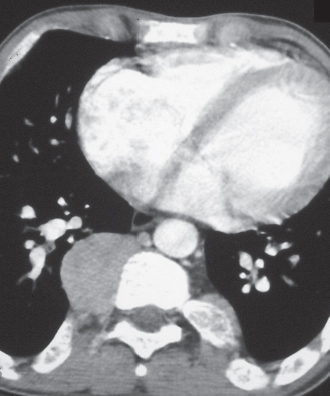
CASE 169 25-year-old man with thalassemia major and profound hemolytic anemia PA (Fig. 169.1) and lateral (Fig. 169.2) chest radiographs demonstrate a well-defined right paravertebral soft-tissue mass (arrow) and expansion of osseous medullary spaces. Note the left upper quadrant metallic surgical clips from a prior splenectomy. Contrast-enhanced chest CT (mediastinal window) (Figs. 169.3, 169.4) shows an ovoid well-defined right paravertebral soft-tissue mass and a smaller left paravertebral mass. Note expansion and trabeculation of the marrow spaces of adjacent ribs. Extramedullary Hematopoiesis • Neurogenic Neoplasm • Lymphadenopathy Fig. 169.1 Fig. 169.2 Fig. 169.3 Fig. 169.4
 Clinical Presentation
 Radiologic Findings
 Diagnosis
 Differential Diagnosis
 Discussion
Background
Related posts:
![]()
Stay updated, free articles. Join our Telegram channel

Full access? Get Clinical Tree


Radiology Key
Fastest Radiology Insight Engine


