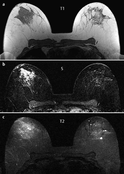
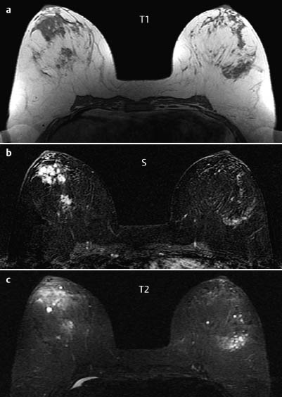
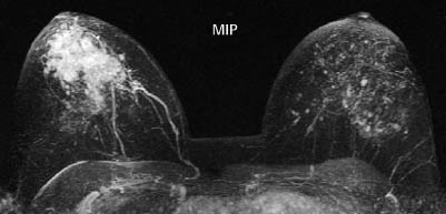

MRM score | Finding | Points |
Shape | dendritic | 1 |
Border | ill-defined | 1 |
CM Distribution | inhomogeneous | 1 |
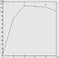
Initial Signal Intensity Increase | strong | 2 |
Post-initial Signal Intensity Character | plateau | 1 |
MRI score (points) |
| 6 |
MRI BI-RADS |
| 5 |
 Preliminary Diagnosis
DCIS, minimally invasive carcinoma, invasive carcinoma.
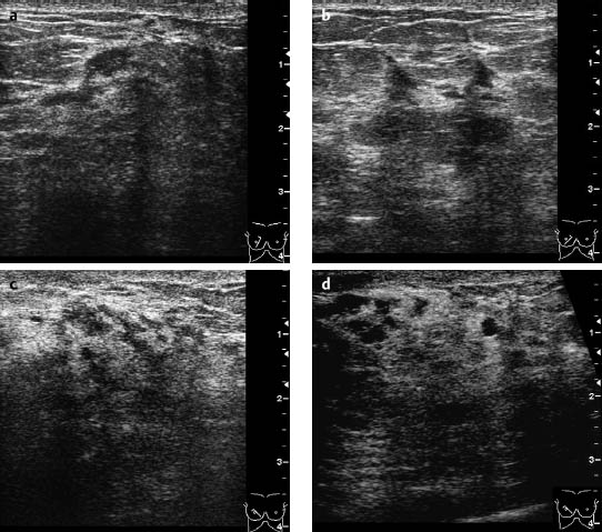
Clinical Findings | right 1 | left 1 |
Ultrasound | right 3 | left 1 |
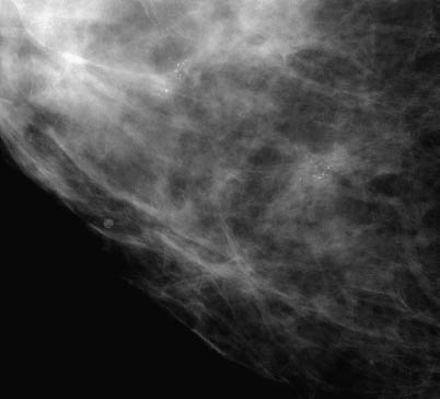
Mammography | right 4 | left 1 |
MR Mammography | right 5 | left 1 |
BI-RADS Total | right 5 | left 1 |
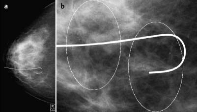
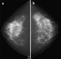
Fig. 17.9a,b Preoperative localization and magnification view (CC).
Procedure
Histopathological evaluation after US-guided core biopsy of the right upper inner quadrant.
Histopathology
Ductal carcinoma in situ.
Further procedure
Preoperative hook-wire localization.
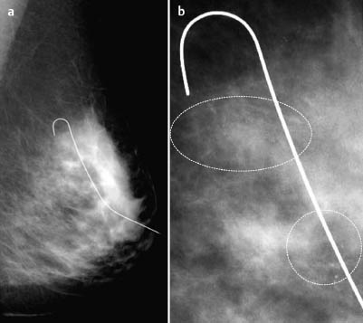
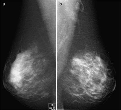
Fig. 17.10a, b Preoperative localization and magnification view (MLO). Both calcification groups are marked with a Homer wire.
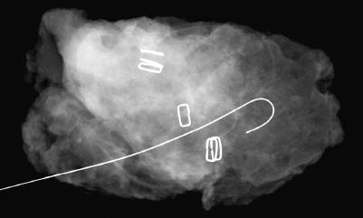
Fig. 17.11 Specimen radiography.
Histology
Extended ductal carcinoma in situ. As expected, no infiltration of the axillary lymph nodes.
DCIS, pN0, G2

Stay updated, free articles. Join our Telegram channel

Full access? Get Clinical Tree


