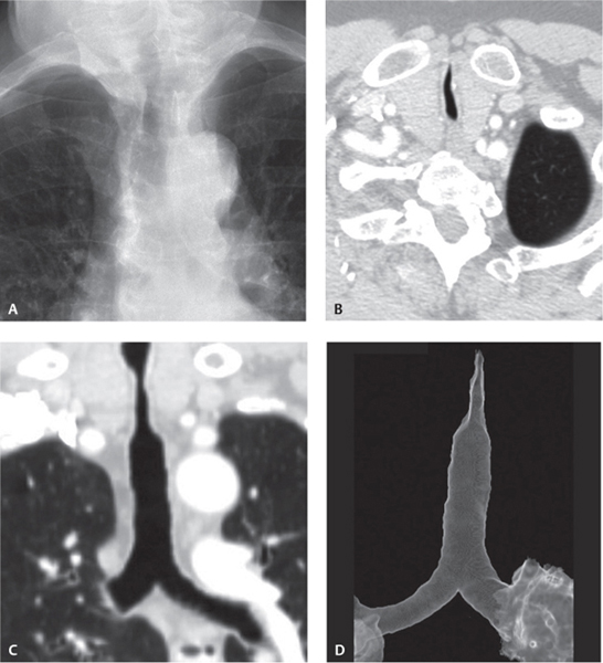
CASE 17 34-year-old man with stridor that developed six months after prolonged tracheostomy Coned-down PA chest radiograph (Fig. 17.1A) demonstrates symmetric narrowing of the tracheal lumen. Chest CT (lung window) with axial and coronal reformation (Figs. 17.1B, 17.1C) demonstrates tracheal narrowing with a reduced coronal diameter and anterior luminal tapering. Surface rendered 3-D image (Fig. 17.1D) of tracheal and central airways shows a tapered area of stenosis in the upper trachea at the level of previous tracheostomy. Fig. 17.1 Tracheal Stenosis; Post-Intubation Injury • Benign and Malignant Tracheal Neoplasm • Wegener Granulomatosis • Amyloidosis Tracheal stenosis is defined as narrowing of the tracheal lumen by more than 10% of its normal diameter. It is a relatively uncommon condition with a frequently insidious onset. Early signs and symptoms may be disregarded or confused with other disorders.
 Clinical Presentation
 Radiologic Findings
 Diagnosis
 Differential Diagnosis
 Discussion
Background
Etiology
Related posts:


Stay updated, free articles. Join our Telegram channel

Full access? Get Clinical Tree


