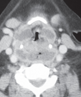
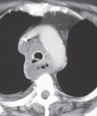
CASE 170 70-year-old woman evaluated for retropharyngeal abscess and chest pain Contrast-enhanced neck CT (soft-tissue window) (Fig. 170.1) demonstrates a complex peripharyngeal fluid collection and a retropharyngeal abscess. Note enhancing borders of the complex fluid collection and air bubbles within the retropharyngeal abscess. Contrast-enhanced chest CT (mediastinal window) (Fig. 170.2) shows a mediastinal fluid collection with enhancing borders and intrinsic air bubbles that surrounds the trachea and esophagus and abuts the visualized thoracic great vessels. This process represented mediastinal extension of the retropharyngeal abscess. Note small right pleural effusion. Mediastinal Abscess • Tuberculous Mediastinitis • Infected Neoplasm Fig. 170.1 Fig. 170.2 (Images courtesy of Diane C. Strollo, MD, University of Pittsburgh Medical Center, Pittsburgh, Pennsylvania.) Acute mediastinal infection is an uncommon but potentially life-threatening inflammatory condition.
 Clinical Presentation
 Radiologic Findings
 Diagnosis
 Differential Diagnosis
 Discussion
Background
Etiology
Related posts:


Stay updated, free articles. Join our Telegram channel

Full access? Get Clinical Tree


