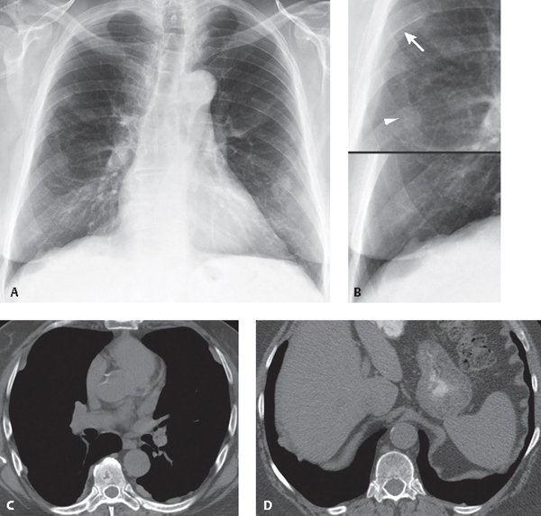
CASE 173 78-year-old man with a history of colon cancer PA chest radiograph (Fig. 173.1A) and coned-down composite views of right lung (Fig. 173.1B) demonstrate multi-focal bilateral nodular opacities. Note discrepant visualization of the lesion borders (Fig. 173.1B) (ill definition of some lesion borders [arrowhead] and sharp margination [arrow] of others) consistent with an extrapulmonary location. Unenhanced chest CT (mediastinal window) (Figs. 173.1C, 173.1D) shows multi-focal discontinuous areas of pleural thickening along the costal and diaphragmatic pleural surfaces, many of which are adjacent to ribs. Fig. 173.1 Pleural Plaques None Four types of benign asbestos-related pleural disease are recognized: non-calcified pleural plaques, calcified pleural plaques, benign asbestos pleural effusion, and diffuse pleural fibrosis (see Table 125.1). Pleural plaques are the macroscopic and radiologic hallmarks of past asbestos exposure and typically develop 15–20 years after initial exposure. Pleural plaques tend to occur adjacent to relatively rigid structures such as the ribs, vertebrae, and the tendinous portion of the diaphragms. Subsequent calcium deposition often occurs in pleural plaques, beginning as fine, punctate flecks that may coalesce over time to form dense streaks or plate-like deposits. Benign asbestos pleural effusions are dose-related manifestations of asbestos exposure that may develop within a shorter latency period than that of other asbestos-related pleural diseases (1–20 years). The effusions are typically unilateral, typically exudates, and sometimes hemorrhagic. The diagnosis is established based on a history of exposure and through exclusion of other etiologies, particularly malignancy. Diffuse pleural thickening and diffuse pleural fibrosis are thought to develop after a previous asbestos-related pleural effusion, involve the visceral pleura, and affect the pleural surface adjacent to the costophrenic angle, a distinguishing feature from pleural plaques.
 Clinical Presentation
 Radiologic Findings

 Diagnosis
 Differential Diagnosis
 Discussion
Background
Etiology
Stay updated, free articles. Join our Telegram channel

Full access? Get Clinical Tree


