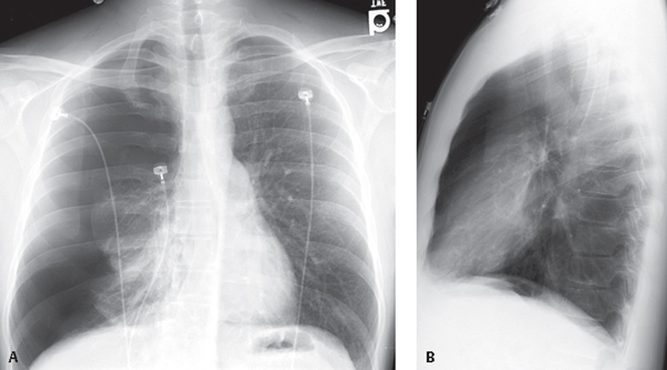
CASE 174 37-year-old man presents to the Emergency Department with pleuritic chest pain and shortness of breath PA (Fig. 174.1A) and lateral (Fig. 174.1B) chest X-rays show a right-sided primary spontaneous pneumothorax with near total collapse of the right lung and contralateral mediastinal displacement. Primary Spontaneous Pneumothorax • Secondary Spontaneous Pneumothorax • Traumatic Pneumothorax Pneumothorax is the presence of air in the pleural space and is a common form of thoracic disease. Fig. 174.1 Pneumothorax may be classified into one of three major categories: traumatic, which can be either iatrogenic (following unsuccessful central venous line or pacemaker-ICD placement, CT-guided or transbronchial biopsy, etc.) or non-iatrogenic (e.g., following blunt or penetrating chest injuries (see Case 84); primary spontaneous, caused by rupture of an air-containing space within (bleb) or immediately deep to the pleura (bulla) (Fig. 174.2); or secondary spontaneous, related to underlying lung disease (e.g., emphysema, chronic interstitial fibrosis or cystic lung disease). Pneumothorax may also develop from pneumomediastinum as a result of air tracking from the mediastinal pleura. Primary spontaneous pneumothorax occurs most often in men (2–15M: 1F) in the third or fourth decade of life and shows a right-sided predilection and strong association with tobacco abuse. Numerous underlying conditions may be associated with secondary spontaneous pneumothorax (Table 174.1), the most common of which is COPD. In some cases, the secondary spontaneous pneumothorax is the first clinical manifestation of the underlying disease. Chest pain and/or dyspnea are the classic clinical symptoms of spontaneous pneumothorax. Breath sounds are decreased or absent on auscultation despite normal or increased resonance on percussion. • Focal areas of apical emphysema or bulla typically not seen • Visceral pleural reflection
 Clinical Presentation
 Radiologic Findings
 Diagnosis
 Differential Diagnosis
 Discussion
Background
Etiology
Clinical Findings
Imaging Findings
Primary Spontaneous Pneumothorax
Chest Radiography
 Vascular markings do not extend beyond lateral border (Fig. 174.1A)
Related posts:


Stay updated, free articles. Join our Telegram channel

Full access? Get Clinical Tree


