Case 18
Indication: Pain in right breast.
History: Unremarkable.
Risk profile: No increased risk.
Age: 55 years.
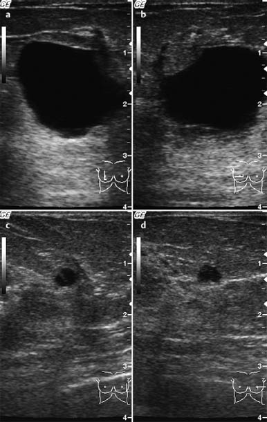
Fig. 18.1 a-d Ultrasound.
Clinical Findings
Resistance of 2 cm diameter in the right breast, above the areola.
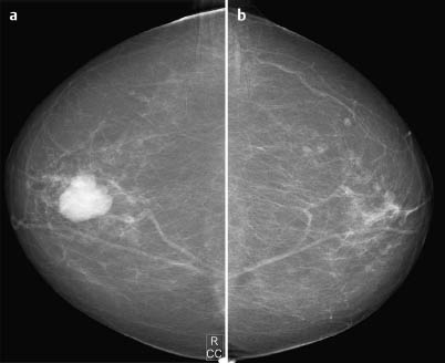
Fig. 18.2a,b Digital mammography, CC view.
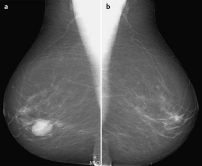
Fig. 18.3a,b Digital mammography, MLO view.
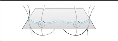
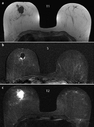
Fig. 18.4a -c Contrast-enhanced MR mammography.
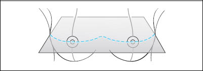
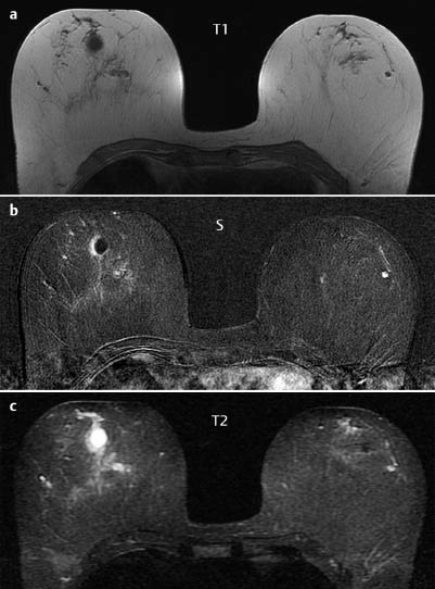
Fig. 18.5a-c Contrast-enhanced MR mammography.
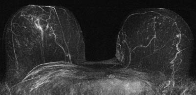
Fig. 18.6 Contrast-enhanced MR mammography. Maximum intensity projection.
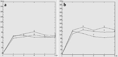
Fig. 18.7a,b Signal-to-time curves of the cyst wall, right breast (a) and of the lesion in the left breast (b).

|
Please characterize ultrasound, mammography and MRI.
What is your preliminary diagnosis?
What are your next steps? |


