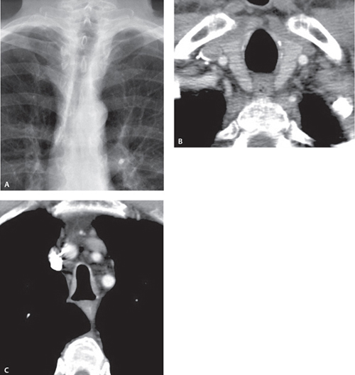
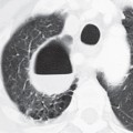
CASE 18 62-year-old man with chronic obstructive pulmonary disease Coned-down PA chest radiograph (Fig. 18.1A) demonstrates narrowing of the tracheal coronal diameter. Contrast-enhanced chest CT (mediastinal window) demonstrates normal tracheal morphology at the level of the thoracic inlet (Fig. 18.1B) and deformity of the tracheal lumen below that level (Fig. 18.1C). The tracheal wall is of normal thickness. The cross-sectional morphology of the tracheal lumen resembles that of a saber sheath. Fig. 18.1 Saber Sheath Trachea • Tracheal Stricture • Relapsing Polychondritis • Amyloidosis • Tracheobronchopathia Osteochondroplastica
 Clinical Presentation
 Radiologic Findings

 Diagnosis
 Differential Diagnosis
Stay updated, free articles. Join our Telegram channel

Full access? Get Clinical Tree


