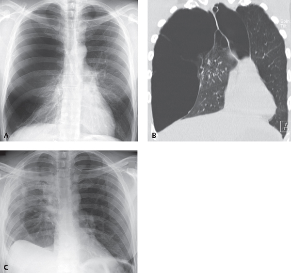
CASE 188 57-year old man with shortness of breath PA chest radiograph (Fig. 188.1A) demonstrates pulmonary hyperinflation with diaphragmatic flattening and increased lucency with giant bullae in both lungs. Compressive changes are noted in the right lower lobe with crowding of bronchovascular structures. Unenhanced chest CT (Fig. 188.1B) shows giant bullae in both lungs, right larger than left, with leftward shift of the mediastinal structures. PA chest radiograph following lung volume reduction surgery (Fig. 188.1C) demonstrates reduction in overall lung volumes, increased upward convexity of the left hemidiaphragm, and improved aeration in the lung parenchyma bilaterally. A subpulmonic effusion obscures the right lateral sulcus and displaces the apex of the diaphragm laterally. Lung Volume Reduction Surgery (LVRS) for Giant Bullous Emphysema Fig. 188.1 (Images courtesy of Anthony D. Cassano, MD, Department of Thoracic Surgery, VCU Medical Center, Richmond, Virginia.) None
 Clinical Presentation
 Radiologic Findings
 Diagnosis

 Differential Diagnosis
 Discussion
Background
Stay updated, free articles. Join our Telegram channel

Full access? Get Clinical Tree


