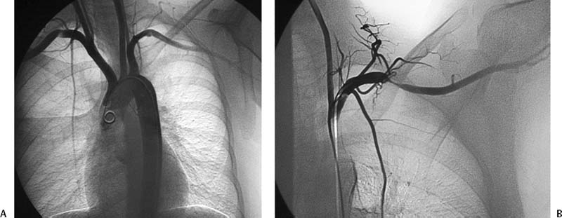
CASE 19 A 35-year-old male presented to the outpatient clinic complaining of sudden onset of a blue middle finger of the left hand. He described a history of crampy muscle pains in both upper extremities. Figure 19-1 Diagnosis of thoracic outlet syndrome with arterial involvement. (A) Arch aortogram with the arms in the neutral position shows aneurysmal dilatation of the left subclavian artery at the level of the thoracic outlet with a linear defect suggesting mild extrinsic compression. The right subclavian artery has a similar linear defect at the thoracic outlet. (B) Selective left subclavian arteriogram with the arm in hyperabduction shows near-complete compression of the subclavian artery at the thoracic outlet. Arch aortogram with the arms in the neutral position (Fig. 19-1A) showed aneurysmal dilatation of the left subclavian artery at the level of the thoracic outlet with a linear defect suggesting mild extrinsic compression. The right subclavian artery had a similar linear defect at the thoracic outlet. Selective left subclavian arteriogram with the arm in hyperabduction (Fig. 19-1B) showed near-complete compression of the subclavian artery at the thoracic outlet. Thoracic outlet syndrome (TOS). The patient underwent transaxillary resection of the left first rib with release of the insertion of the anterior scalene muscle.
Clinical Presentation
Radiologic Studies
Diagnosis
Treatment
Equipment
Discussion
Background
Related posts:


Stay updated, free articles. Join our Telegram channel

Full access? Get Clinical Tree


