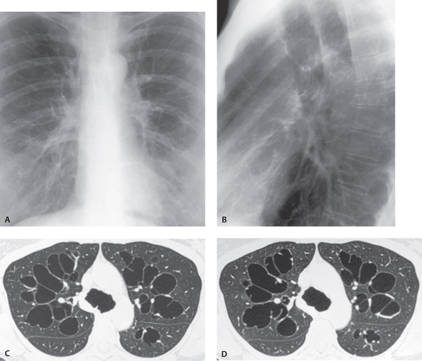
CASE 19 38-year-old woman with recurrent respiratory infections Coned-down PA (Fig. 19.1A) and lateral (Fig. 19.1B) chest radiographs demonstrate enlargement of the tracheal lumen and bilateral central thin-walled pulmonary cystic lesions. HRCT (lung window) (Figs. 19.1C, 19.1D) demonstrates marked tracheal enlargement involving the origins of the mainstem bronchi. Note the corrugated contour of the tracheal wall and bilateral branching thin-walled cystic lesions located centrally in the lung, consistent with bronchiectasis. Fig. 19.1 Tracheobronchomegaly; Mounier-Kuhn Syndrome • Bronchiectasis • Williams-Campbell Syndrome • Chronic Airway Inflammation/Infection with Tracheobronchomalacia • Allergic Bronchopulmonary Fungal Disease Tracheobronchomegaly or Mounier-Kuhn syndrome is a rare condition also known as tracheal diverticulosis and tracheobronchiectasis. The etiology of tracheobronchomegaly is unknown. Some authors have postulated a congenital etiology
 Clinical Presentation
 Radiologic Findings
 Diagnosis
 Differential Diagnosis
 Discussion
Background
Etiology
![]()
Stay updated, free articles. Join our Telegram channel


Full access? Get Clinical Tree


