MRM score | Finding | Points |
Shape | irregular | 1 |
Border | ill-defined | 1 |
CM Distribution | dendritic | 1 |
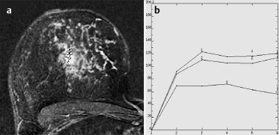
Initial Signal Intensity Increase | strong | 2 |
Post-initial Signal Intensity Character | plateau | 1 |
MRI score (points) |
| 6 |
MRI BI-RADS |
| 5 |
 Preliminary Diagnosis
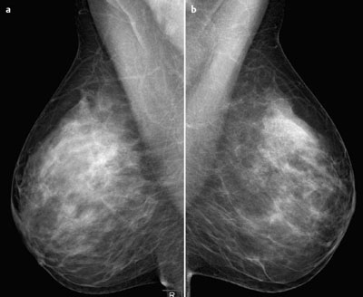
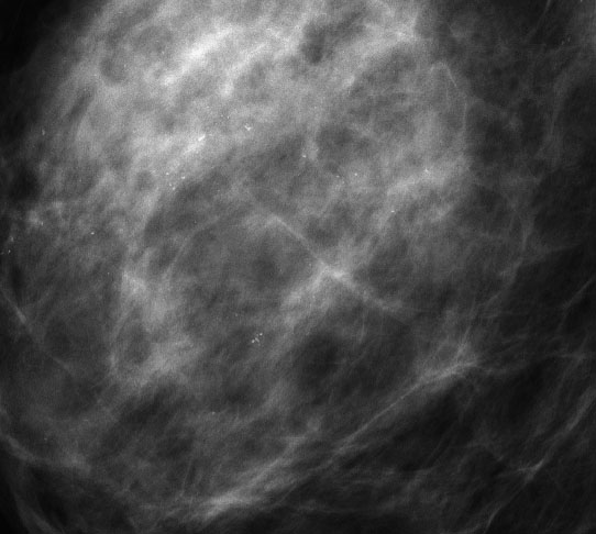
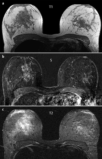
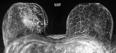
Extensive intraductal, possibly partially microinvasive tumor of the right breast.
Differential Diagnosis
Segmental adenosis, inflammation (both very unlikely).
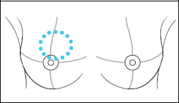
Clinical Findings | right 3 | left 1 |
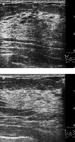
Ultrasound | right 3 | left 1 |
Mammography | right 5 | left 1 |
MR Mammography | right 5 | left 1 |
BI-RADS Total | right 5 | left 1 |
Procedure
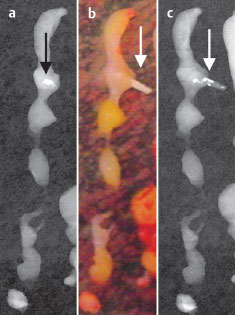
Stereotactic vacuum core biopsy of the right breast to obtain histological confirmation of preliminary diagnosis. Compression with the probe of one of the tissue samples retrieved in the area of a calcification, visible here, produced “blackhead”-like emissions from the core. This is evidence of the presence of intraductal tumor.
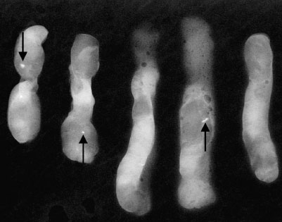
Fig. 2.7 Specimen radiography of some of the core biopsies with positive findings of microcalcifications (arrows).
Fig. 2.8a–c Specimen of a single core biopsy before and after compression of the tissue including the microcalcification. Photography of the necrotic comedo tissue (arrow) after compression.
Histology of the specimen
Comedo-type DCIS, Grade 2.
Histology of the right breast
Tricentric intraductal breast carcinoma in the central part of the right breast including the nipple (see the linear enhancement in MRI reaching the nipple region).
pTis (Paget disease), pN0 (sentinel Node 0/2), G2, R1 (upper inner quadrant), VNP110 points.
Stay updated, free articles. Join our Telegram channel

Full access? Get Clinical Tree


