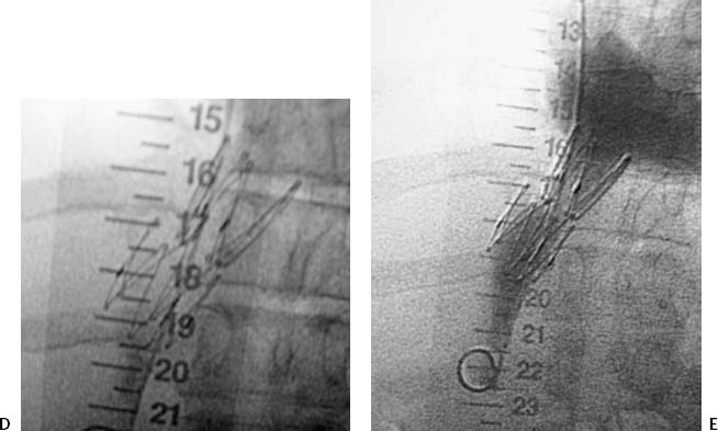
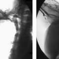
CASE 2 A 47-year-old female had received a liver transplant 8 months before presenting to our vascular and interventional radiology section with massive leg swelling and pain. Figure 2-1 Angioplasty and stenting of symptomatic inferior vena caval stenosis. (A) Digital subtraction venogram shows focal stenosis of the intrahepatic inferior vena cava. (B) Fluoroscopic image shows balloon angioplasty of the stenosis. (C) Postangioplasty venogram shows significant residual stenosis. (D) Fluoroscopic image shows deployed Z-stent. (E) Poststenting venogram shows good flow through the stent without residual stenosis. The right internal jugular vein was catheterized using sonographic guidance. A pigtail catheter was advanced into the inferior vena cava. An inferior vena cavogram was performed revealing a high-grade stenosis of the inferior vena cava at the level of the upper surgical anastomosis (Fig. 2-1A). Pressure measurements obtained across the stenosis revealed a gradient of 26 mm Hg. Post-transplant inferior vena caval stenosis. The diagnostic catheter was exchanged over a rigid guidewire (Amplatz Super Stiff, Boston Scientific, Natick, Massachusetts) for an 18- × 20-mm angioplasty balloon catheter (XXL balloon dilation catheter, Boston Scientific, Natick, Massachusetts) that was used to dilate the lesion (Fig. 2-1B). Repeat cavography revealed minimal improvement (Fig. 2-1C), and the pressure gradient across the narrowed segment remained at 21 mm Hg. Due to the elastic recoil of the lesion, we elected to attempt stent insertion.
Clinical Presentation
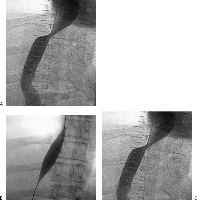
Radiologic Studies
Cavography
Diagnosis
Treatment
Related posts:


Stay updated, free articles. Join our Telegram channel

Full access? Get Clinical Tree


