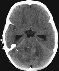
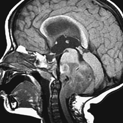
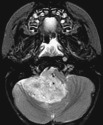
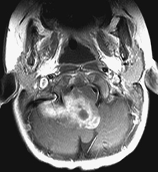
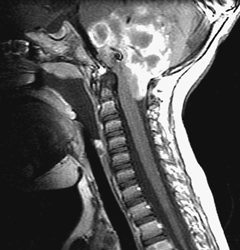
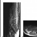
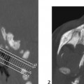
CASE 2 A 2-year-old child presents with early morning vomiting, headaches, progressive seclusion, irritability, and recent torticollis. Figure 2A Figure 2B Figure 2C Figure 2D Figure 2E Nonenhanced CT demonstrates hydrocephalus and a mass in the central posterior fossa. This mass is ill-demarcated and heterogeneous with hypo-, iso-, and hyperdense portions and with punctiform calcifications (Fig. 2A). The fourth ventricular lumen is not seen. Sagittal T1- (Fig. 2B) and axial T2-weighted MR imaging (Fig. 2C) exhibits inhomogeneity with a large infiltration of the brain stem, whereas the vermis appears pushed backward instead. Although centered on the fourth ventricle, the mass shows cisternal extensions through the ventricular foramina into the upper cervical canal and the right cerebellopontine cistern. Contrast enhancement (Fig. 2D) is relatively mild and irregular and confirms the poor demarcation of the lesion. Spinal imaging (Fig. 2E) is important, as ependymomas tend to generate drop metastases in the CSF spaces. Posterior fossa ependymoma
Clinical Presentation
Radiologic Findings
Diagnosis
Differential Diagnosis
Related posts:


Stay updated, free articles. Join our Telegram channel

Full access? Get Clinical Tree


