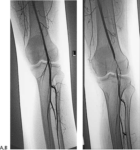
CASE 21 A 32-year-old male presented to the outpatient clinic with intermittent left lower extremity calf claudication. Figure 21-1 Diagnosis of popliteal artery entrapment syndrome. (A) Selected left lower extremity arteriogram with the foot in passive dorsiflexion shows a patent popliteal artery with three-vessel runoff to the lower leg. (B) Repeat selected arteriogram with the foot in active plantar flexion shows extrinsic compression causing complete occlusion of the left popliteal artery. The right common femoral artery was punctured using the Seldinger technique, and a 5-French (F) sheath was inserted. The left external iliac artery was selected. An arteriogram (Fig. 21-1A) with the left foot in passive dorsiflexion showed a patent popliteal artery with three-vessel runoff to the lower leg. Repeat selected left leg arteriogram (Fig. 21-1B) with the foot in active plantar flexion showed extrinsic compression causing complete occlusion of the left popliteal artery with distal reconstitution by collateral vessels. Intermittent obstruction of the left popliteal artery resulting from popliteal artery entrapment syndrome (PAES). The patient underwent surgical decompression and has been symptom-free for 8 months. Puncture needle 5F vascular sheath 5F pigtail catheter 0.035” conventional soft-tipped guidewire Contrast material PAES describes extrinsic compression of the popliteal artery caused by abnormal position of this vessel in relation to the surrounding structures. PAES has been divided into five types (Table 21-1
Clinical Presentation
Radiologic Studies
Angiography
Diagnosis
Treatment
Equipment
Discussion
Background
![]()
Stay updated, free articles. Join our Telegram channel


Full access? Get Clinical Tree


