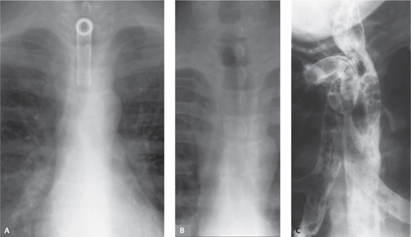
CASE 21 37-year-old man with cough after swallowing Coned-down PA chest radiograph (Fig. 21.1A) demonstrates a tracheostomy tube in place; the tube was removed 6 weeks later. Coned-down AP chest radiograph (Fig. 21.1B) obtained one week after tracheostomy tube removal demonstrates air distension of the esophagus that overlies the normal tracheal air column. Coned-done oblique (RPO) radiograph (Fig. 21.1C) obtained during Gastrografin swallow demonstrates a fistulous communication between the posterior wall of the trachea and the esophagus at the level of a replaced tracheostomy tube. Contrast material opacifies the esophagus, trachea, tracheostomy stoma, and both mainstem bronchi (Fig. 21.1C). Tracheoesophageal Fistula; Complication of Prolonged Tracheostomy Tube Placement None Fig. 21.1
 Clinical Presentation
 Radiologic Findings
 Diagnosis
 Differential Diagnosis
Related posts:


Stay updated, free articles. Join our Telegram channel

Full access? Get Clinical Tree


