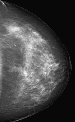
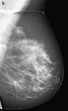
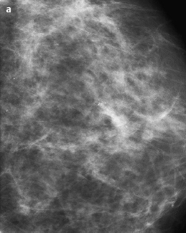
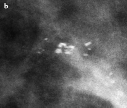
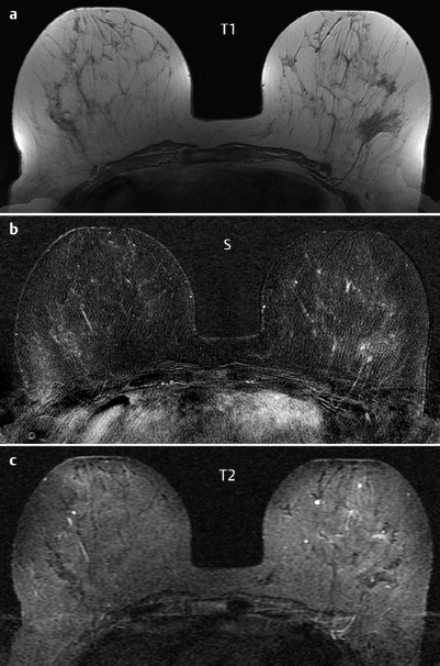
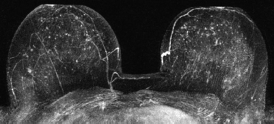
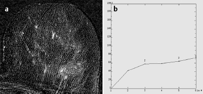
Case 22 Indication: Screening. History: Unremarkable. Risk profile: No increased risk. Age: 55 years. Fig. 22.1 a Digital mammography, CC view. Fig. 22.1 b Digital mammography, MLO view. Normal. No pathological findings (not shown). Fig. 22.2a Magnification view (CC). Fig. 22.2 b Zoomed view of the magnification view. Fig. 22.3a–c Contrast-enhanced MRI of the breasts. Fig. 22.4 Contrast-enhanced MR mammography. Maximum intensity projection. Fig. 22.5a,b Signal-to-time curve of the Y-shaped enhancement. Please characterize ultrasound, mammography, and MRI findings. What is your preliminary diagnosis? What are your next steps? Sonography showed multiple cysts (maximum diameter 5 mm) in both breasts, and otherwise normal echo patterns. There were no suspicious findings in the central part of the left breast or elsewhere. US BI-RADS right 2/left 2. Imaging showed fibroglandular parenchyma, ACR type 2. There were polymorphous microcalcifications (round and linear, no V-or Y-shaped particles) in the central part of the left breast in a linear or segmental orientation. Mammograms showed no masses or densities and no architectural distortions. BI-RADS right l[no figure]/left 4. PGMI is defined only for bilateral mammography. MRI depicted a Y-shaped enhancement in the center of the left breast, most likely in the same location as the microcalcifications detected in mammography (Fig. 22.6). MRI Artifact Category: 1 MRI Density Type: 1 Fig. 22.6 Y-shaped enhancement in the left breast.
Clinical Findings
Ultrasound

Ultrasound
Mammography
MR Mammography

MRM score | Finding | Points |
Shape | dendritic | 1 |
Border | well-defined | 0 |
CM Distribution | homogenous | 0 |
Initial Signal Intensity Increase | moderate | 1 |
Post-initial Signal Intensity Character | increasing | 0 |
MRI score (points) |
| 2 |
MRI BI-RADS |
| 2 |
 Preliminary Diagnosis
DCIS.
Differential Diagnosis
Papilloma, adenosis, ductal hyperplasia, focal mastitis.
Clinical Findings | right 1 | left 1 |
Ultrasound | right 2 | left 2 |
Mammography | right 1 | left 4 |
MR Mammography | right 1 | left 2 |
BI-RADS Total | right 2 | left 4 |
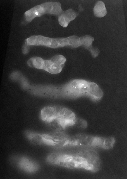
Fig. 22.7 Radiogram of some of the specimens.
Histology (vacuum core biopsy)
Intraductal papillomatosis.

Stay updated, free articles. Join our Telegram channel

Full access? Get Clinical Tree


