23

Case 23


Indication: Screening.


History: Unremarkable.


Risk profile: No increased risk.


Age: 56 years.


image


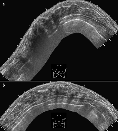
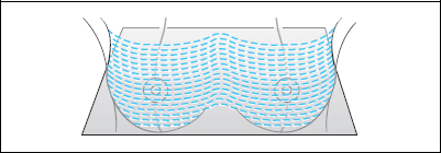
Fig. 23.1 a,b Sonography. Panoramic view of right and left breasts.


image


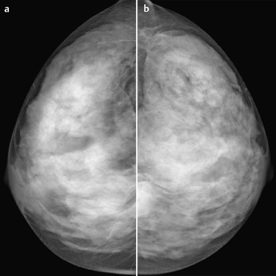
Fig. 23.2a,b Digital mammography, CC view.


image


Clinical Findings


Resistant parenchyma in the upper outer quadrants of both breasts, known and unchanged over a number of years. No circumscribed mass.


image


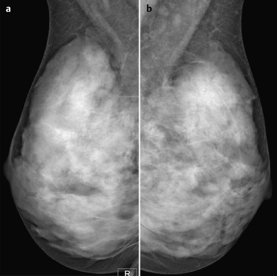
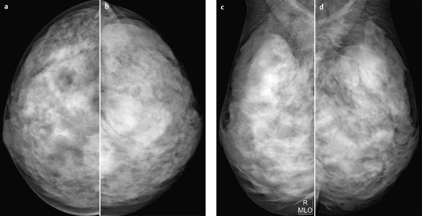
Fig. 23.3a,b Digital mammography, MLO view.


image


image


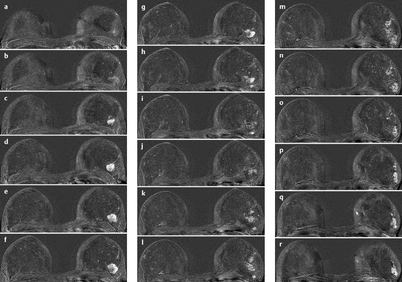
Fig. 23.4a–r Contrast-enhanced MR mammography. 18 consecutive subtraction images, cranial to caudal.


image


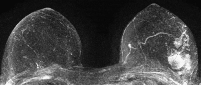
Fig. 23.5 Contrast-enhanced MR mammography. Maximum intensity projection.


image


Please characterize ultrasound, mammography and MRI.


What is your preliminary diagnosis?


What are your next steps?


The indication for examination was a screening situation. Clinical examination did not demonstrate any difference between the right and the left breasts, even though the MRI findings suggest that this patient has a large mass, which one would expect to be palpable, in her left breast.


Ultrasound


Panoramic views of the upper quadrants of both breasts are shown. Comparing both sides, suspect acoustic shadowing is in evidence on the right, while there are no notable ultrasound findings in the left breast. US BI-RADS right 3/left 2.


Mammography


Imaging showed bilaterally symmetric extremely dense parenchyma, ACR type 4. Considering these limiting conditions, no masses or densities could be identified. Mammograms demonstrated no architectural distortions or microcalcifications. BI-RADS right 1/left 1. PGMI: CC view P; MLO view M (inframammary fold not correctly depicted, pectoralis muscle does not reach nipple line).


MR Mammography


All presented subtraction images showed pathological multiple enhancement in the outer quadrants of the left breast. The morphology of the enhancing areas was highly pathological and their T2 signal was also suspect (intermediate signal, not shown). Signal-time curves also indicated malignancy (strong initial signal increase, washout; not shown).


MRI Artifact Category: 1


MRI Density Type: 2


MRM score


Finding


Points


Shape


irregular


1


Border


ill-defined


1


CM Distribution


rim sign


2


Initial Signal Intensity Increase


strong


2


Post-initial Signal Intensity Character


wash out


2


MRI score (points)


8


MRI BI-RADS


5


image Preliminary Diagnosis


Extended invasive carcinoma. No differential Diagnosis.


BI-RADS Categorization


Clinical Findings


right 2


left 2


Ultrasound


right


left 2


Mammography


right 1


left 1


MR Mammography


right 1


left 5


BI-RADS Total


right 1


left 5


Procedure


Histopathological evaluation of the extensive enhancing areas in the lateral parts of the left breast with MR-guided core biopsy, because ultrasound did not provide clearly corresponding data. MR-guided vacuum biopsy in the upper outer quadrant of the left breast (11 gauge, 12 specimens). Additionally, MR-guided core biopsy in the lower outer quadrant of the left breast (16G, 8 specimens spaced 1 cm apart) to verify multicentricity.


Histopathology (all specimens)


Tubular breast cancer grade 1 -2.


image


Fig. 23.6a-d Digital mammography in 2 views from screening examination a year earlier, with identical findings.


Sep 3, 2016 | Posted by in GENERAL RADIOLOGY | Comments Off on 23

Full access? Get Clinical Tree
Get Clinical Tree app for offline access