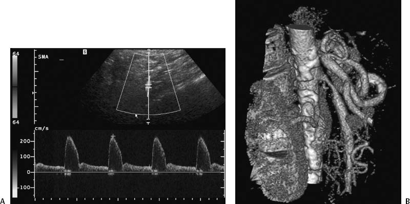
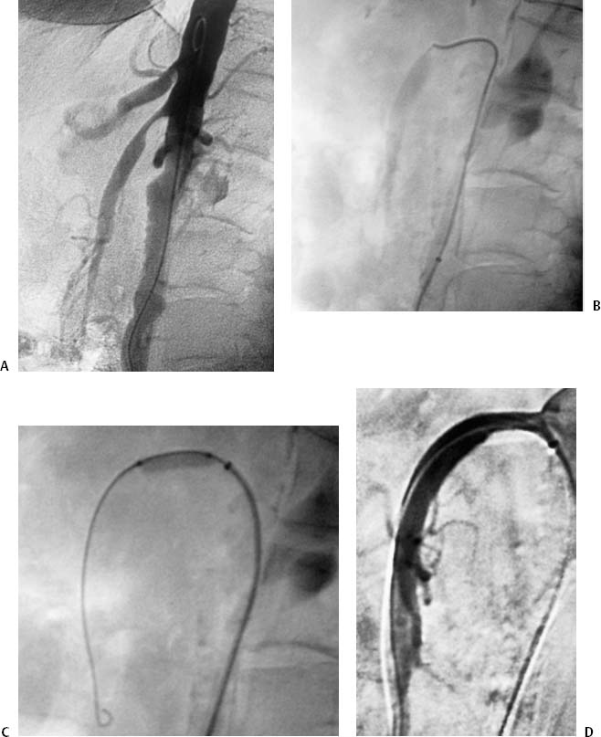
CASE 23 A 58-year-old female who had lost 20 pounds over the past year presented with complaints of abdominal pain after eating and “food fear.” Figure 23-1 Noninvasive evaluation of superior mesenteric stenosis. (A) Doppler ultrasound of SMA reveals an elevated velocity of 233 cm/s. (B) CTA shows high-grade stenoses of both the celiac and SMA. An abdominal ultrasound was performed. Doppler interrogation of the superior mesenteric artery (SMA) revealed a velocity of 233 cm/s (Fig. 23-1A). CTA showed high-grade stenoses of both the celiac artery and the SMA (Fig. 23-1B). The right common femoral artery was catheterized using the Seldinger technique, and a 6-French (F) Balkan sheath was placed. A pigtail catheter was advanced into the abdominal aorta and lateral aortography was performed showing moderate-to-high grade stenoses of both the superior mesenteric and celiac arteries (Fig. 23-2A). Figure 23-2 (A) Lateral aortogram confirms CTA findings of high-grade mesenteric stenoses. (B) Lateral fluoroscopic image shows positioning of stent at stenosis. (C) Lateral fluoroscopic image shows balloon deployment of stent. (D) Digital subtraction angiogram shows alleviation of the SMA stenosis. Celiac and SMA stenoses with symptoms of chronic mesenteric ischemia. Puncture needle or micropuncture kit 5F and 6F guiding sheath 5F pigtail and endhole catheters 0.035” and 0.014” angioplasty balloons, wires, and balloon-expandable stents (4 to 6 mm) Contrast material Heparin Inflation syringe
Clinical Presentation
Radiologic Studies
Doppler Ultrasound
CT Angiography (CTA)
Angiogram
Diagnosis
Treatment
Equipment
Mesenteric Angioplasty and Stent Insertion
Related posts:


Stay updated, free articles. Join our Telegram channel

Full access? Get Clinical Tree


