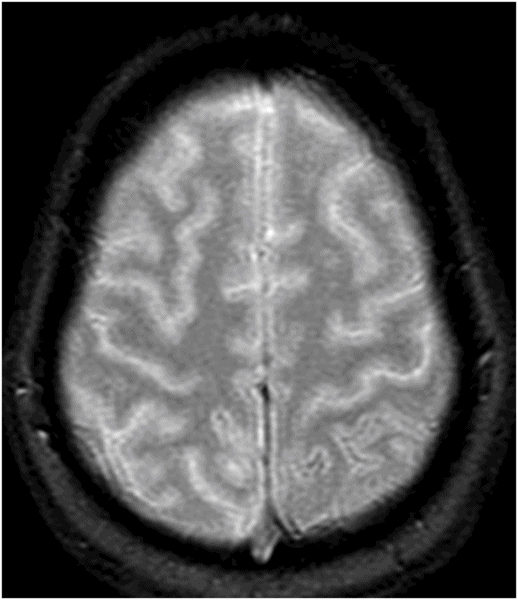
Axial GRE image through the same level.
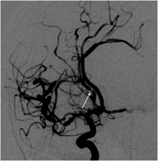
Catheter angiogram of the left internal carotid artery, oblique projection.
Reversible Cerebral Vasoconstriction Syndrome
Primary Diagnosis
Reversible cerebral vasoconstriction syndrome
Differential Diagnoses
Cerebral vasculitis (primary angiitis of CNS)
Systemic lupus
Non-aneurysmal subarachnoid hemorrhage (dural fistula, trauma, arteriovenous malformation)
Aneurysmal subarachnoid hemorrhage
Migraines
Imaging Findings
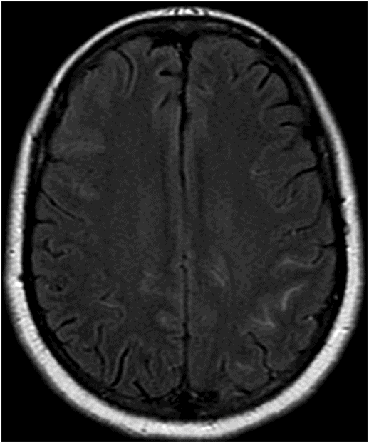
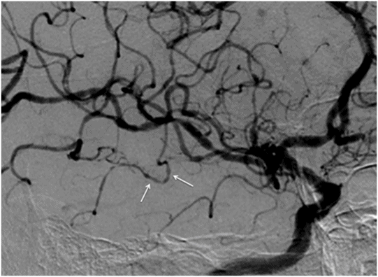
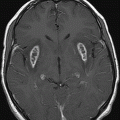
Fig. 25.1: Axial head CT through the level of central sulcus demonstrated subtle subarachnoid hemorrhage in the left parietal region, including the postcentral sulcus. Fig. 25.2: Axial FLAIR image through the level of central sulcus demonstrated subtle subarachnoid hemorrhage in the left parietal region, including the postcentral sulcus. Fig. 25.3: Axial GRE image through the level of central sulcus confirmed subarachnoid hemorrhage in the left postcentral sulcus. Fig. 25.4: Catheter angiogram of the left internal carotid artery on the lateral projection demonstrated multifocal beading of one of the left M3 branches (arrows). Fig. 25.5: Catheter angiogram of the left internal carotid artery on the oblique projection demonstrated focal narrowing of the left pericallosal artery (arrow).
Discussion
Typical clinical presentation (recurrent acute thunderclap headache in a young female patient) associated with typical imaging findings (subarachnoid hemorrhage [SAH] close to the vertex, focal beading, and narrowing of the arteries) are consistent with the diagnosis of reversible cerebral vasoconstriction syndrome (RCVS).
Central nervous system vasculitides, particularly primary angiitis of CNS (PACNS), can have a similar angiographic appearance but are more frequently associated with a gradual symptomatic onset. Moreover, thunderclap headaches have never been reported in patients with PACNS. These patients demonstrate inflammatory CSF abnormalities whereas MRI scans are abnormal in most cases of PACNS, showing small deep or superficial infarcts of different ages, with or without white matter signal abnormalities. In addition, complications including posterior reversible encephalopathy syndrome (PRES), infarctions, and hemorrhages typically occur in watershed zones in PACNS, whereas in RCVS, hemorrhages can be distributed over watershed zones.
Aneurysmal SAH is more diffuse and typically involves the basilar cisterns whereas SAH is typically minimal and involves the cerebral convexity, overlying a few cortical sulci in RCVS. Delayed vasospasm in aneurysmal SAH may be a differential consideration on angiography; however, it is usually long segmental and in close proximity to the bleeding site. In contrast, RCVS has diffuse, short segmental vasoconstriction. Cerebral angiography would be diagnostic for both aneurysms and entities such as arteriovenous malformation or dural fistula. Migrainous headache does not have a specific imaging abnormality.
Reversible cerebral vasoconstriction syndrome refers to a group of disorders characterized by reversible, multifocal cerebral arterial vasoconstriction, most commonly manifesting as severe acute, usually recurrent thunderclap headaches, with or without other acute neurologic symptoms including nausea, vomiting, photophobia, confusion, and blurred vision. This entity has a female predominance (3:1) and most patients are middle-aged. Typically, the headaches recur for one to two weeks. Ischemic and hemorrhagic stroke are the major complications (7–54%). Other complications include PRES (9–14%), brain edema (38%), cortical SAH (up to 34%), or subdural hemorrhages (2%). Ischemic events predominantly occur during the second week, later than the hemorrhagic events that tend to occur in the first week. These complications can arise after complete resolution of headache as well.
This syndrome may be spontaneous, or primary in approximately one-third of patients. It may be secondary and associated most commonly with use of vasoactive substances (e.g., recreational drugs such as cannabis or cocaine, or drugs such as SSRIs, nasal decongestants, pseudoephedrine, and occasionally immunosuppressant or cytotoxic agents) or with a postpartum state (about 9%). Additional noted associations have been with tumors such as pheochromocytoma, glomus tumors, or bronchial carcinoid tumor, cervical artery or aortic dissection, unruptured intracranial aneurysm, fibromuscular dysplasia, postcarotid endarterectomy, erythropoietin, intracranial hypotension, intracranial hemorrhage, spinal subdural hematoma, neurosurgery, head trauma, and miscellaneous states such as porphyria.
Pathophysiology of RCVS remains unclear: possible causes include transient dysfunctional regulation of cerebral vascular tone that leads to segmental vasoconstriction and vasodilatation in small vessels. This triggers thunderclap headache by abruptly stretching vessel walls. Oxidative stress, sympathetic overactivity, aberrant sympathetic response of the vessels, endothelial dysfunction, and alterations in biochemical and immunologic factors regulating vascular tone, have been suggested as potential causes. Histologic studies have showed no evidence of arterial inflammation or infection.
Non-contrast CT is often negative but may show small cortical SAH in 20% (presumably from minor leaks or rupture of surface vessels), with or without parenchymal hemorrhage. Typical imaging findings include convexity SAH, intracerebral hemorrhage, and intracerebral edema. Convexity SAH is usually mild, and limited to a few cerebral sulci, and appears as hyperintense on FLAIR and hypointense on GRE images. Intracerebral hemorrhage is of variable volume and is usually solitary and lobar. Infarction is typically in the arterial borderzone areas, often between the posterior cerebral artery and anterior circulation. Computed tomography or MR angiography may be normal (10%) but may show diffuse segmental arterial vasoconstriction (90%). Occasionally, if the first study was negative, second imaging studies obtained one to two weeks later may show positive findings.
Digital subtraction angiography is critical for diagnosis and demonstrates diffuse, multifocal, segmental narrowing in the large and medium-sized arteries that is frequently bilateral. Occasional dilated segments may give a string-of-beads appearance. If the angiogram is performed early, within a week of symptomatic onset, it may be completely normal, even with presence of SAH, cerebral infarction, and intracerebral hemorrhage. Angiogram should be repeated after several days if the clinical suspicion is strong, and often follow-up angiogram is positive. By definition, the abnormalities are transient, and should show complete resolution on repeat imaging one to three months later. Cerebrospinal fluid analysis is usually normal or near normal (protein concentrations < 100 mg/dl, < 15 white blood cells per μl).
Reversible cerebral vasoconstriction syndrome should be considered an emergent condition and treatment includes discontinuation of the offending agent and use of vasodilators such as calcium channel blockers (e.g., nimodipine). The disease has an overall favorable long-term prognosis, influenced by the occurrence of stroke. In a series, 71% of patients in the long term had no evidence of disability and 29% had only minor disability. Most patients with strokes gradually improve for several weeks, and few have residual deficits and <5% develop life-threatening forms with several strokes and massive brain edema.


Stay updated, free articles. Join our Telegram channel

Full access? Get Clinical Tree


