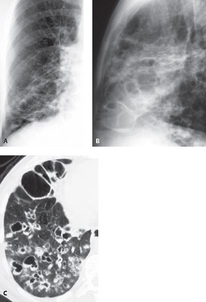
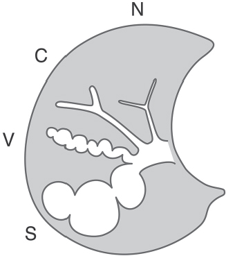
CASE 25 36-year-old man with chronic productive cough and recurrent pneumonia Coned-down PA (Fig. 25.1A) and lateral (Fig. 25.1B) chest radiographs demonstrate central coarse linear and interstitial opacities with ring-like and cystic morphology and a bronchial distribution affecting the middle lobe and right lower lobe. Unenhanced chest CT (lung window) (Fig. 25.1C) targeted at the lower right lung demonstrates areas of moderate and severe bronchiectasis with bronchial wall thickening. Note the varied morphology of the bronchiectatic airways, including cylindrical, varicoid, and saccular (cystic) forms (Figs. 25.1C, 25.2). Multiple areas of mucoid impaction and endoluminal air-fluid levels are seen in the right lower lobe (Fig. 25.1C). Fig. 25.1 Fig. 25.2 Artist’s illustration depicts a normal bronchus (N) and the three grades of severity of bronchiectasis: cylindrical (C), varicose (V), and saccular (cystic) (S). Bronchiectasis with Mucous Plugging; Secondary Superinfection None Bronchiectasis is defined as chronic irreversible bronchial dilatation and may be secondary to a variety of inflammatory and destructive bronchial wall disorders. It is typically graded according to its severity as mild, moderate, and severe forms, respectively termed cylindrical, varicose, and saccular (cystic) (Fig. 25.2). Cylindrical bronchiectasis is characterized by mild bronchial dilatation with preservation of bronchial morphology (Figs. 25.2, 25.3, 25.4, 25.6). Varicose bronchiectasis is characterized by areas of bronchial dilatation alternating with foci of luminal constriction that result in a beaded bronchial morphology (Figs. 25.1C, 25.2, 25.4, 25.6). The most severe form of bronchiectasis is characterized by cystic or saccular bronchial dilatation measuring over 1.0 cm in diameter (Figs. 25.1A, 25.1B, 25.1C, 25.2, 25.7). All forms of bronchiectasis are associated with bronchial wall thickening. The underlying mechanism behind most forms of bronchiectasis is bronchial wall injury. Etiologies include childhood viral and bacterial infections, cystic fibrosis, ciliary dyskinesia syndromes, immunodeficiency disorders, allergic bronchopulmonary fungal disease, and lung and bone marrow transplantation. Pulmonary fibrosis may cause bronchiectasis by retraction of peribronchial fibrous tissue (traction bronchiectasis) and is usually a localized process (e.g., tuberculosis, sarcoidosis, pulmonary fibrosis, radiation therapy).
 Clinical Presentation
 Radiologic Findings
 Diagnosis
 Differential Diagnosis
 Discussion
Background
Etiology
Clinical Findings
Related posts:
![]()
Stay updated, free articles. Join our Telegram channel

Full access? Get Clinical Tree


Radiology Key
Fastest Radiology Insight Engine


