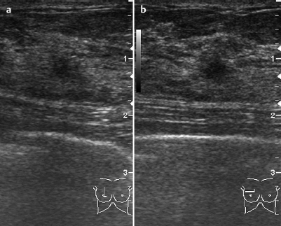
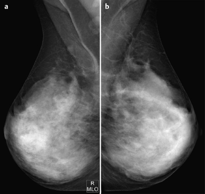
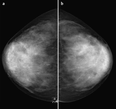
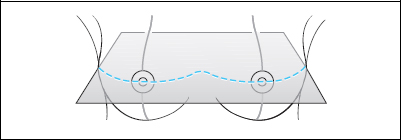
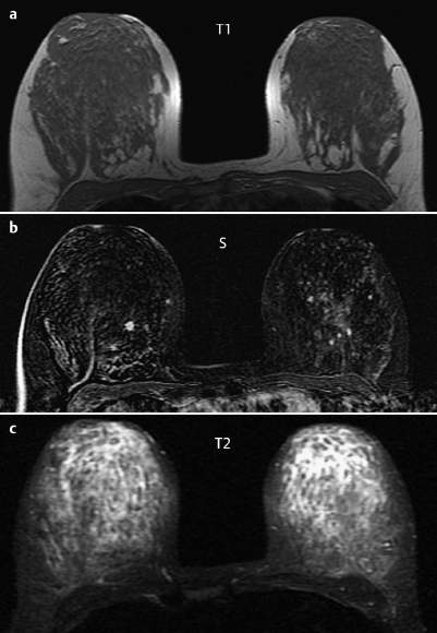
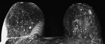
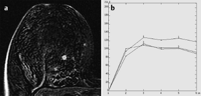
Case 26 Indication: Screening mammography. History: Unremarkable. Risk profile: No increased risk. Age: 49 years. Fig. 26.1 a,b Ultrasound Fig. 26.2a,b Digital mammography, MLO view. No abnormalities. Fig. 26.3a,b Digital mammography, CC view. Fig. 26.4a–c Contrast-enhanced MRI of the breasts. Fig. 26.5 Contrast-enhanced MR mammography. Maximum intensity projection. Fig. 26.6a,b Signal-to-time curves. Please characterize ultrasound, mammography, and MRI findings. What is your preliminary diagnosis? What are your next steps? This case presents the imaging studies of an asymptomatic woman in a screening situation. Retrospectively, in view of the MRI findings, an ill-defined hypoechoic lesion of 5 mm diameter with irregular borders and indeterminate distal echo behavior was depicted by sonography. There was no architectural distortion. US BI-RADS right 3. Mammography showed bilaterally symmetric, extremely dense parenchyma, ACR type 4. Particularly in the inner quadrants of the right breast, there were no suspicious densities or lesions and no microcalcifications. No architectural distortion (BI-RADS right 1/left 1). PGMI: CC view P; MLO view G (inframammary fold, axillary skin fold). Between the inner quadrants of the right breast, there was an ill-defined, spiculated lesion of approx. 5 mm diameter. This lesion demonstrated ring enhancement, a strong initial signal increase of 140%, and a postinitial plateau. Signal in T2-weighted imaging was indeterminate. No further suspect lesions. MRI Artifact Category: 2 MRI Density Type: 2
Clinical Findings

Ultrasound
Mammography
MR Mammography
MRM score | Finding | Points |
Shape | round | 0 |
Border | spiculated | 1 |
CM Distribution | ring | 2 |
Initial Signal Intensity Increase | strong | 2 |
Post-initial Signal Intensity Character | plateau | 1 |
MRI score (points) |
| 6 |
MRI BI-RADS |
| 5 |
 Preliminary Diagnosis
Carcinoma.
Differential Diagnosis
Focal adenosis.
Clinical Findings | right 1 | left 1 |
Ultrasound | right 3 | left 1 |
Mammography | right 1 | left 1 |
MR Mammography | right 5 | left 1 |
BI-RADS Total | right 5 | left 1 |
Consideration
Does the sonographically depicted lesion correspond exactly with the enhancing area in MR mammography? Since the imaging evidence was not conclusive on this point, MR-guided investigation of the lesion was recommended.
Procedure
MS-guided vacuum biopsy of the lesion in the center of the right breast.
Histopathology of the biopsy specimen, right breast
Adenosis.
Diagnosis
Adenosis.
Stay updated, free articles. Join our Telegram channel

Full access? Get Clinical Tree


