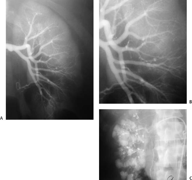
CASE 27 A 54-year-old male immigrant from Eastern Europe presented with complaints of fever, abdominal pain, weight loss, and malaise. He was hypertensive, and proteinuria was found on urinalysis. Figure 27-1 PAN. (A) Selective left renal arteriogram shows numerous microaneurysms throughout the kidney. (B) Magnified image of A. (C) Venous phase of selective right renal arteriogram in a different patient with PAN shows innumerable microaneurysms. The patient was referred to interventional radiology for arteriography. The right common femoral artery was punctured using the Seldinger technique and a 5-French (F) sheath was inserted. A pigtail catheter was advanced into the abdominal aorta and an aortogram was performed (not shown). The left renal artery was selectively catheterized using an RC-1 (Boston Scientific, Natick, Massachusetts) catheter, and a selective renal arteriogram was performed, which revealed numerous microaneurysms throughout the kidney (Fig. 27-1). Biopsy of the skin was then performed, showing polyarteritis nodosa (PAN). The patient was treated with high-dose steroids and cytotoxic drugs. Puncture needle 5F vascular sheath 5F pigtail, RC-1, RIM, and Visceral Selective-1 catheters (Boston Scientific, Natick, Massachusetts) Contrast material
Clinical Presentation
Radiologic Studies
Diagnosis
Treatment
Equipment
Discussion
Background
Related posts:


Stay updated, free articles. Join our Telegram channel

Full access? Get Clinical Tree


