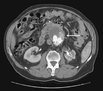
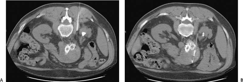
CASE 28 A 62-year-old male had undergone aortic endograft repair of a 5.5-cm abdominal aortic aneurysm 1 year before coming to us. A routine CT exam obtained 6 months after stent graft insertion was obtained (Fig. 28-1). Figure 28-1 Endoleak. CTA shows perfusion of excluded aneurysm sac (arrow). Note atrophic left kidney. CTA showed contrast pooling in the aneurysm sac adjacent to the stent graft. This is termed an endoleak. Endoleaks represent a failure of aneurysm exclusion and may be associated with enlargement and rupture. This was thought to be a Type 2 leak. In our hospital, in the absence of aneurysm enlargement, Type 2 endoleaks are closely followed. This patient was followed expectantly, and a CT exam 1 year after repair showed the aneurysm had enlarged to 6 cm in diameter. The patient was then referred to interventional radiology for endoleak embolization. Type 2 endoleak with aneurysm enlargement. The patient was given preprocedural antibiotics. Using CT guidance, a translumbar puncture using a 22-gauge needle was made into the aneurysm sac (Fig. 28-2A). Pulsatile blood return confirmed location within the leak. A pressure measurement revealed a sac pressure of 87 mm Hg. A 0.018-inch guidewire was inserted into the sac, and the needle was removed (Fig. 28-2B). The patient was then transferred to the interventional radiology suite. A 5-French (F) catheter was advanced into the sac and an initial angiogram was obtained (Fig. 28-3A). The catheter was then manipulated into the inferior mesenteric artery (IMA) (Fig. 28-3B) and an angiogram was obtained. Multiple coils were deployed into the origin of the IMA (Fig. 28-3C). Gelfoam (Pharmacia and Upjohn, Kalamazoo, MI) was deployed into the aneurysm sac. A follow-up CTA performed 3 days after the procedure (Fig. 28-4) showed contrast stasis in the aneurysm sac from the procedure indicating successful occlusion of the leak.
Clinical Presentation
Radiologic Studies
CT Angiography (CTA)
WHAT IS THE ABNORMALITY?
WHAT IS APPROPRIATE FOLLOW-UP?
Diagnosis
Treatment
CT-Guided Translumbar Puncture
Embolization
Follow-up CTA
Discussion
Background
Related posts:


Stay updated, free articles. Join our Telegram channel

Full access? Get Clinical Tree


