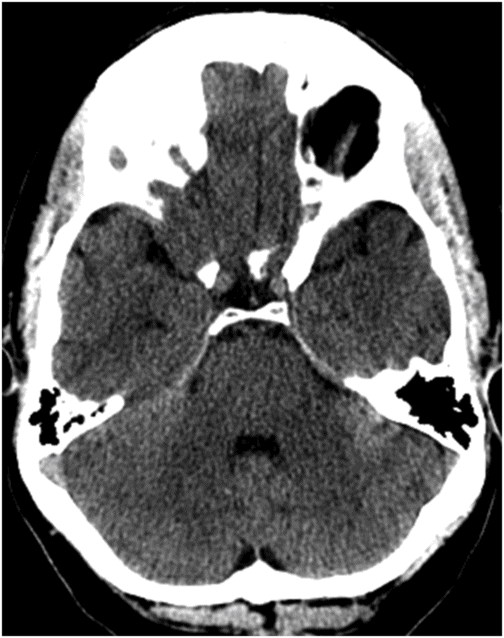
Axial non-contrast CT scan of the head through the level of the pons.

Axial non-contrast CT scan of head through the level of the pons, at a slightly superior level.
Perimesencephalic Subarachnoid Hemorrhage
Primary Diagnosis
Perimesencephalic subarachnoid hemorrhage
Differential Diagnoses
Posterior fossa AVM
Craniovertebral junction/upper cervical AVM
Bleeding from vascular tumors
Imaging Findings
Fig. 29.1: Axial non-contrast CT scan of the head through the level of the pons demonstrated thin, slit-like subarachnoid hemorrhage in the prepontine cistern, limited immediately anterior to the pons. Note there is no blood in the fourth ventricle. Fig. 29.2: Axial non-contrast CT scan of head through the level of the pons, at a slightly superior level, did not demonstrate blood in the prepontine or perimesencephalic cistern. Blood was absent from the suprasellar cistern, basal aspect of the sylvian fissures, and anterior interhemispheric fissure. The patient was further evaluated with CT angiogram of the head (not shown) that was negative.
Discussion
Minute subarachnoid hemorrhage (SAH) limited immediately anterior to the pons with no extension to the perimesencephalic cistern, suprasellar cistern, or the basal aspect of the sylvian or interhemispheric fissures, in a 47-year-old patient, is suggestive of perimesencephalic subarachnoid hemorrhage (PMSAH). The non-contrast CT scan and the CT angiogram did not reveal any mass in the posterior fossa or any vascular malformation, thus eliminating consideration of vascular tumor.
Perimesencephalic subarachnoid hemorrhage accounts for nearly 10% of non-traumatic SAH and approximately one-third of non-traumatic non-aneurysmal SAH. Unlike aneurysmal SAH, PMSAH does not have any known sex predilection. Perimesencephalic subarachnoid hemorrhage is characterized by the presence of hemorrhage that is centered only in the anterior perimesencephalic cistern (anterior to midbrain and pons) with variable extension to interpeduncular, ambient, and quadrigeminal cisterns. Minimal blood may extend to the suprasellar cistern, inferior aspect of the anterior interhemispheric cistern, and basal portion of the sylvian fissure but may not extend to the distal or more superior aspects of these cisterns. Subtle extension of hemorrhage into the occipital horn is also possible in PMSAH. Definitive PMSAH diagnosis is valid only on non-contrast CT scan of the head obtained within three days of symptom onset. Patients meeting these imaging criteria have a very favorable prognosis as no aneurysm is seen in up to 95% of these patients. There are no delayed complications associated with PMSAH such as rebleeding, or vasospasm, unlike other causes of SAH. The cause of PMSAH is venous; however, in up to 5% of cases, it may be related to aneurysm rupture of the vertebrobasilar system. Other uncommon causes are vascular malformation in the posterior fossa or in the craniovertebral junction.
Patients meeting PMSAH criteria should be evaluated with CT angiogram of the head to rule out other possibilities. If other causes are found, appropriate management is necessary. If the CT angiogram is negative, a digital subtraction angiogram (DSA) may not be required for further evaluation, as DSA is usually negative.
Stay updated, free articles. Join our Telegram channel

Full access? Get Clinical Tree


