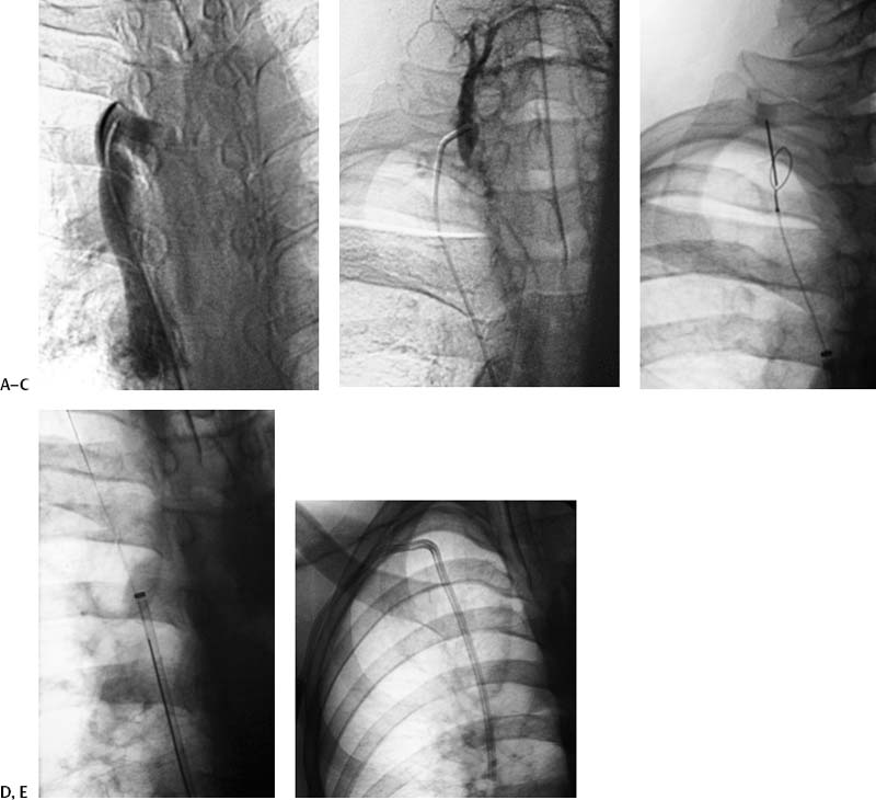
CASE 29 A 45-year-old male who had end-stage renal disease requiring hemodialysis had a history of numerous central line infections causing thromboses and subsequent obstruction. He was referred to interventional radiology for tunneled dialysis catheter insertion. Figure 29-1 Venous recanalization. (A) Venogram shows occlusion of superior vena cava at the level of the azygous. (B) Venogram shows recanalization of the small vein. (C) Fluoroscopic image shows 21-gauge needle advanced through the loop of the snare. (D) Fluoroscopic image shows captured guidewire pulled into the femoral vein sheath. (E) Final fluoroscopic image shows right-sided catheter placed via the recanalized vein. Sonographic evaluation of the neck revealed complete thrombosis of both internal and external jugular veins and occlusions of both subclavian veins. Complete upper central venous occlusion. The chest, neck, and right groin were cleansed, and antibiotics were administered prophylactically (ceftizoxime sodium, 1 g IV). The right common femoral vein was punctured and a guiding 9-French (F) vascular introducer sheath was advanced into the superior vena cava (SVC). A venogram confirmed occlusion of the SVC at the level of the azygous vein (Fig. 29-1A). A 180-cm rigid hydrophilic guidewire and hydrophilic coated 5F catheter were advanced into the brachiocephalic veins. The catheter and guidewire were used to engage a collateral vein in the neck (Fig. 29-1B). Sonography was performed to choose a suitable puncture site away from overlying arteries. The catheter was exchanged for a 10-mm snare, which was opened in the vessel and used as a target. The loop portion was punctured with fluoroscopic guidance using a 21-gauge needle (Fig. 29-1C), the needle was exchanged for a 0.018” guidewire, and the snare was closed and used to pull the guidewire into the SVC (Fig. 29-1D).
Clinical Presentation
Radiologic Studies
WHAT ARE THE OPTIONS FOR TUNNELED CATHETER PLACEMENT AT THIS POINT?
Diagnosis
Treatment
Related posts:


Stay updated, free articles. Join our Telegram channel

Full access? Get Clinical Tree


