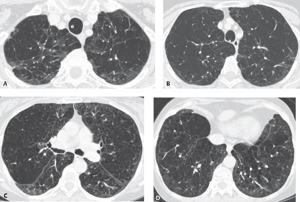
CASE 29 72-year-old woman with exertional dyspnea Unenhanced chest CT (lung window) (Figs. 29.1A, 29.1B, 29.1C, 29.1D) demonstrates bilateral decreased attenuation throughout both lungs with simplification of lung architecture and diffuse hypovascularity. Panacinar (Panlobular) Emphysema • α-1-Antiprotease (Antitrypsin) Deficiency Fig. 29.1 Panacinar (syn. panlobular) emphysema affects each acinus in its entirety and all acini within the secondary pulmonary lobule (Fig. 29.2, right). It is the characteristic finding in patients with α-1-antiprotease deficiency. While panacinar emphysema may involve the lung diffusely, predominant lower lung involvement is characteristic. This is in contradistinction to proximal acinar emphysema, which predominantly involves the upper lungs. Panacinar emphysema is less common than proximal acinar (centrilobular) emphysema. The familial form of panacinar emphysema is seen in association with α-1-antiprotease deficiency. Panacinar emphysema also occurs as a result of intravenous drug abuse (e.g., talc, Ritalin [Novartis, New York]) and in segments of lung affected by congenital bronchial atresia.
 Clinical Presentation
 Radiologic Findings
 Diagnosis
 Differential Diagnosis
 Discussion
Background
Etiology
Clinical Findings
Related posts:


Stay updated, free articles. Join our Telegram channel

Full access? Get Clinical Tree


