3

Case 3


Indication: Screening.


History: Unremarkable.


Risk profile: Postmenopausal breast cancer in mother, premenopausal in an aunt, postmenopausal in a second cousin.


Age: 40 years.


image


Clinical Findings


No findings.


Sonography


No findings (not shown).


image


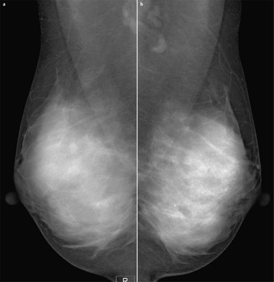
Fig. 3.1 a,b Digital mammography of both breasts, MLO view.


image


image


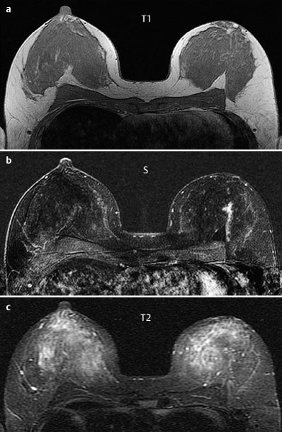
Fig. 3.2a–c Contrast-enhanced MRI of the breasts.


image


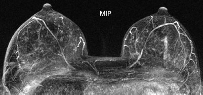
Fig. 3.3 Contrast-enhanced MR mammography. Maximum intensity projection.


image


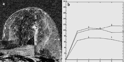
Fig. 3.4a,b Signal-to-time curves.


image


Please characterize mammography and MRI findings.


What is your preliminary diagnosis?


What are your next steps?


These are the imaging studies of a younger woman presenting for screening with a high risk profile due to three relatives having breast cancer.


Ultrasound


Ultrasound showed no suspicious changes (not shown).


Mammography


Bilaterally symmetric, extremely dense parenchymal pattern, ACR type 4. Within the limitations of these conditions, there were no abnormal findings in the left breast central area-where MRI findings are significant-or anywhere else. There is no hyperdensity, no mass, no architectural distortion. No microcalcifications corresponding to the linear enhancement observed in MRI (BI-RADS right 1/left 1). PGMI: MLO view P.


MR Mammography


There is linear and partially dendritic enhancement, likely representing a milk duct, visible in the central sections of the left breast. Signal-time curves are nonspecific. No further observations.


MRI Artifact Category: 1


MRI Density Type: 1


MRM score


Finding


Points


Shape


linear


1


Border


ill-defined


1


CM Distribution


homogeneous


0


Initial Signal Intensity Increase


strong


2


Post-initial Signal Intensity Character


plateau


1


MRI score (points)


5


MRI BI-RADS


4


image Preliminary Diagnosis


DCIS.


Differential Diagnosis


Segmental papillomatosis, inflammation of the milk ducts.


BI-RADS Categorization


Clinical Findings


right 1


left 1


Ultrasound


right 1


left 1


Mammography


right 1


left 1


MR Mammography


right 1


left 4


BI-RADS Total


right 1


left 4


Procedure


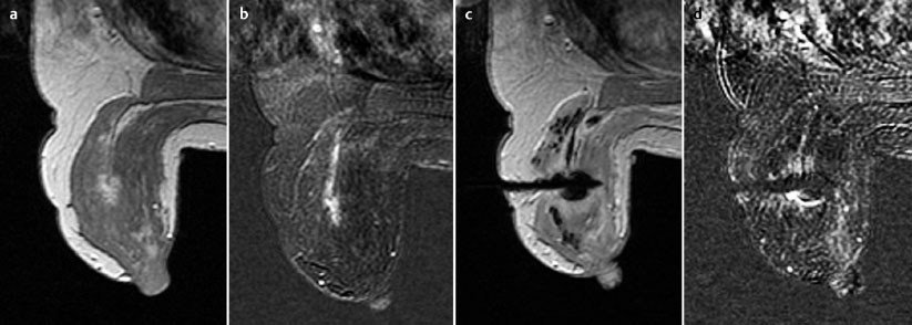
Histological verification of the suspicious findings in MRI, preferably with MR-guided percutaneous vacuum biopsy to avoid open biopsy.


image


Fig. 3.5a–d Documentation of the MR-guided vacuum biopsy: T1-weighted precontrast image of the left breast positioned within the stereotactic device (a). Precise reproducibility of the linear enhancement in subtraction image (b). Position of the biopsy needle after removal of 20 core biopsies (11 gauge) (c). Documentation of parts of the enhancing area in a second contrast-enhanced MRI sequence (subtraction image) (d).


Sep 3, 2016 | Posted by in GENERAL RADIOLOGY | Comments Off on 3

Full access? Get Clinical Tree
Get Clinical Tree app for offline access