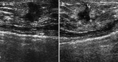
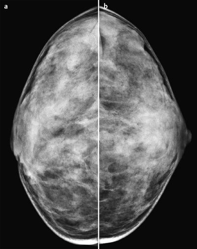
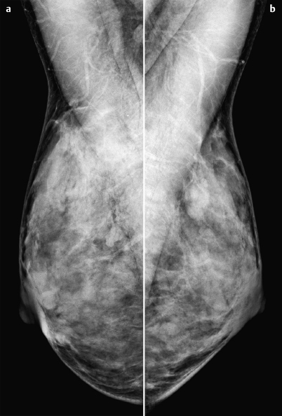
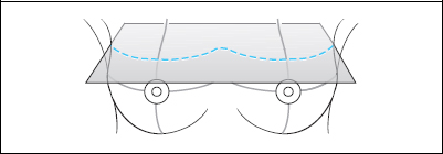
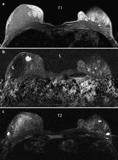
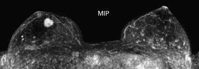
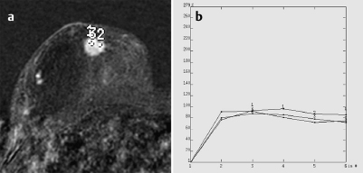
Case 31 Indication: Palpable mass in the right breast, discovered 2 months previously. History: Unremarkable. Risk profile: No increased risk. Age: 42 years. Fig. 31.1 a,b Ultrasound image of the region of the resistance. Dense lump 2 cm diameter in the right breast above the nipple. Fig. 31.2a,b Digital mammography, CC view. Fig. 31.3a,b Digital mammography, MLO view. Fig. 31.4a–c Contrast-enhanced MRI of the breasts. Fig. 31.5 Contrast-enhanced MR mammography. Maximum intensity proiection. Fig. 31.6a,b Signal-to-time curves. Please characterize ultrasound, mammography, and MRI findings. What is your preliminary diagnosis? What are your next steps? Corresponding to the known lump, ultrasound showed a lobulated and well-defined lesion with a markedly hyperechoic margin and accompanying distortion of the surrounding tissue. US BI-RADS 5 right. Mammograms showed bilaterally symmetric, extremely dense parenchyma, ACR type 4. Due to the limitations of these conditions, no abnormalities were visible in the area of the palpable mass or elsewhere. There were no architectural distortions or calcifications (BI-RADS right 1/left 1). PGMI: CC view P; MLO view G (inframammary fold). MRI demonstrated a round, hypervascularized lesion about 1 cm in diameter and with partially ill-defined borders in the right breast, cranial to the nipple. This lesion corresponds to the clinical and sonographic findings in the same location and had an intermediate signal in T2 imaging. The nonspecific signal-time curves showed an initial increase of about 70% followed by a plateau. There was also a cyst in the left breast and further spotty enhancing foci in both breasts (see maximum intensity projection, Fig. 31.5). MRI Artifact Category: 1 MRI Density Type: 2
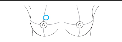
Clinical Findings

Ultrasound
Mammography
MR Mammography
MRM score | Finding | Points |
Shape | round | 0 |
border | ill-defined | 1 |
CM Distribution | inhomogeneous | 1 |
Initial Signal Intensity Increase | moderate | 1 |
Post-initial Signal Intensity Character | plateau | 1 |
MRI score (points) |
| 4 |
MRI BI-RADS |
| 4 |
 Preliminary Diagnosis
Breast cancer.
Clinical Findings | right 3 | left 1 |
Ultrasound | right 5 | left 1 |
Mammography | right 1 | left 1 |
MR Mammography | right 4 | left 2 |
BI-RADS Total | right 5 | left 2 |
Procedure
Histopathological analysis of the lump, preferably by US-guided percutaneous core biopsy.
Performed step (departing from guidelines)
At the patient’s request, open biopsy was performed to remove the mass without primary core biopsy analysis.
Histology
Invasive lobular carcinoma, 12 mm.
ILCpTIc, pN0, G2.
Stay updated, free articles. Join our Telegram channel

Full access? Get Clinical Tree


