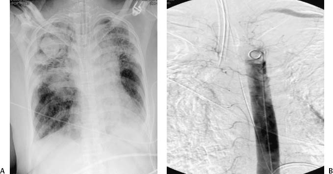
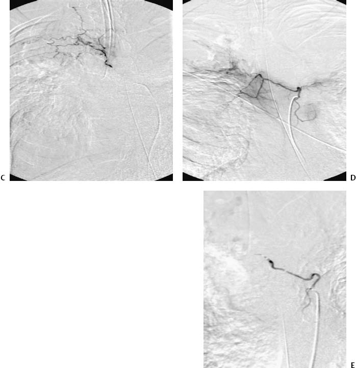
CASE 31 A 45-year-old male with a history of pulmonary tuberculosis presented to the emergency department with severe hemoptysis. Bronchoscopy showed active bleeding within the left main bronchus, limiting further evaluation. Figure 31-1 Diagnosis and treatment of a patient with massive hemoptysis. (A) Chest radiograph shows bilateral interstitial lung disease with predominantly right-sided cavitation and interstitial lung disease. (B) Descending aortogram shows the level of a right-sided bronchial artery. (C) Selected bronchial angiography shows increased vascularity in the distribution of a right bronchial artery but no active extravasation of contrast material. (D) Selected angiography of a second bronchial artery more distal shows a common trunk supplying the right and left sides. (E) Selected bronchial angiography after embolization using polyvinyl alcohol particles shows cessation of flow. Both bronchial arteries were embolized. Chest radiograph showed bilateral interstitial lung disease with predominantly right-sided cavitation and interstitial lung disease. The right common femoral artery was punctured using the Seldinger technique, and a 5-French (F) sheath was inserted. A descending aortogram showed the level of a right-sided bronchial artery. The bronchial arteries were selected using a Cobra 2 catheter (Boston Scientific, Natick, Massachusetts), and angiography was performed showing increased vascularity in the distribution a right bronchial artery but no active extravasation of contrast material. A second bronchial artery supplied both the right and left sides, and increased vascularity to the right upper lobe was again seen. Bronchial artery rupture caused by tuberculosis. A 3F microcatheter was advanced through the Cobra catheter (Boston Scientific, Natick, Massachusetts) into both bronchial arteries. Angiography again revealed increased vascularity in this distribution. Embolization was performed by injection of 500 to 700 micron particles until stasis of flow was achieved. 5F Cobra catheter (Boston Scientific, Natick, MA) Standard 0.035-” guidewire 3F microcatheter 0.014-” microwire 500 to 700 micron polyvinyl alcohol particles Contrast material
Clinical Presentation
Radiologic Studies
Radiography
Angiography
Diagnosis
Treatment
Superselective Embolization
Equipment
Discussion
Background
Related posts:


Stay updated, free articles. Join our Telegram channel

Full access? Get Clinical Tree


