31 Osteopenic Diseases in the Hands
Other than focal osteoporosis induced by inactivity or specific diseases of the hand, a number of systemic diseases causing osteopenic remodeling are also manifested in the hand. These include generalized osteoporosis, osteomalacia, the various forms of hyperparathyroidism, renal osteopathy, and neoplastic diseases. Further differentiation is based largely on laboratory results. Important parameters are calcium and phosphate levels in serum and urine, alkaline phosphatase, vitamin D, and parathyroid hormone in serum. Histologic analysis of an iliac-crest biopsy is only occasionally indicated for differential diagnosis of osteopathy. A common radiographic characteristic of osteopenic skeletal diseases is the increased radiolucency of the bone. For early and complete determination of the finest structural changes in the compact and cancellous bone, radiographic examinations should be performed with mammography films and/ or a magnifying technique. For early diagnosis, several osteodensitometric procedures are available.
Anatomy and Pathoanatomy
The completely differentiated lamellar bones consist of (from outside to inside) periosteum, compact bone, cancellous bone, and bone marrow. Regardless of the cellular and architectural structure, biochemically the following components of the osseous substance are differentiated:
The osteoid is an organic bone matrix and consists largely of collagen fibers. These proteins constitute about 35% of the weight of the bone.
The remaining 65% is made up of mineralized bone substance consisting largely of hydroxyapatite crystals, but also calcium phosphate, calcium carbonate, magnesium phosphate, and others.
Bone metabolism is controlled by a complex regulatory system in which hormones and vitamins determine the production and resorption of osseous components:
Somatotropin (STH) from the adenohypophysis promotes bone growth.
The growth-inhibiting effect of pararenal and sex hormones plays only a minor role in bone metabolism.
Parathyroid hormone (normal value in serum 1.5–6.5 pmol/l) produced in the parathyroid glands activates the osteoclasts to reabsorb bone and thereby increases the serum calcium level.
Calcitonin produced by the parafollicular cells of the thyroid gland (normal value in serum 100 ng/l) has the opposite function. It activates the osteoblasts, thereby indirectly lowering the serum calcium level by increased mineralization of the interstitial substance.
Vitamin D promotes the resorption of calcium from the intestine and the mineralization of the osteoid with hydroxyapatite. For the full effect of provitamin D supplied in food to be exerted, it has to be biochemically modified in three steps: The ring of the provitamin D is cleaved in the skin under the influence of ultraviolet light; then double hydroxylation to 1,25-dihydroxycholecalciferol (= calcitriol; normal value in serum 75–175 pmol/l for adults and 100–250 pmol/l for children) takes place in the liver and the kidneys.
The possible etiologies of osteopenic diseases are listed in Table 31.1 .
Disease Entities
Osteoporosis
Pathoanatomy and Clinical Symptoms
There is reduced bone mass resulting from an imbalance between bone production and resorption, with resorption prevailing. Both cortical and cancellous bone structure are rarefied in favor of increased fatty bone marrow. The density of mineral salts and the histologic structure of the remaining bone are normal. Clinically, osteoporosis often causes fractures, especially of the vertebrae, the neck of the femur, and the distal radius.
Generalized osteoporosis is differentiated from the regional/localized form:
The prototype of generalized osteoporosis is postmenopausal/senile involutional osteoporosis. It affects bones that are largely cancellous, like vertebrae, the pelvis, and ribs, more than the short and long tubular bones, like the distal radius. The incidence of radius fractures, however, also increases considerably even in early menopause. Further causes are endocrinologic diseases (diabetes mellitus, Cushing disease, hyperparathyroidism, acromegaly, hyperthyroidism, and estrogen deficit), pregnancy, nutritive-toxic damage (alcoholism, hepatopathies, malnourishment with protein deficiency, and hypovitaminosis C), neoplastic diseases (leukemias, multiple myeloma, and diffuse bone metastases), and hematologic diseases (hemophilia, sickle-cell anemia, thalassemia, and Christmas disease). Paraplegia or genetic defects (osteogenesis imperfecta, Turner and Klinefelter syndromes, hypophosphatasia, homocystinuria, alcaptonuria, mucopolysaccharidoses, and Gaucher disease) are relatively infrequent causes of osteoporosis. Iatrogenic causes are therapies with corticoids, phenytoin, and heparin (more than 15 000 IU daily).
Regional osteoporosis is manifested primarily in the bones of the extremities, particularly para-articular. Etiology is listed in Table 31.2 .
|
Diagnostic Imaging
Radiography
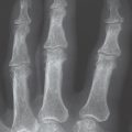
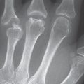
The classic radiographic signs of osteoporosis, which can only be seen in advanced stages, are summarized in Table 31.3 and Fig. 31.1 .
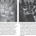
Measurement of the cortical thickness (Barnett-Nordin’s metacarpal index) provides assessment of the bone mass. This involves determining the thickness of the cortical layer on the radial and ulnar sides of the shafts of metacarpals II or III and adding them (a 1 + a 2). The sum is then divided by the entire thickness b of the bone ( Fig. 31.3 ). The quotient (a 1 + a 2)/b is normally greater than 0.5. In osteoporosis, it is below 0.43.
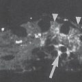
Structural bone changes do not appear with lowactivity osteoporosis. Aside from an increase in radiolucency and a decrease in the thickness of the cortical bone layer, highly active, aggressive osteoporosis leads to subchondral, endosteal, periosteal, and intracortical resorption with tunneling of the compact bone.
Procedures for Measuring Bone Density
On radiographs, osteoporosis can be recognized only when the loss of bone mineral substance and bone structure has reached a level of at least 30–35%. For this reason, the following methods of early diagnosis of osteopenia have been developed:
In single-photon absorptiometry (SPA), an iodine-125 (125I) radiation source (27.5 keV) is moved over the distal forearm in a grid pattern. Attenuation profiles are measured that are equivalent to the mineral content of the bone and are calculated as linear values (g/cm) or mineral content per unit area (g/cm2). SPA has proved useful in large epidemiologic studies and is widely performed.
Dual photon absorptiometry (DPA) is the two-energyvariant of SPA. As emitter substance, the gadolinium isotope 153Gd is usually used. This produces radiation with energies of 44 keV and 100 keV. Assessment of the intensities and the difference of the absorption values at different energies eliminates errors caused by surrounding soft tissues. However, the radiation exposure is considerably higher, and the results have an error rate of 2.5%.
|


A disadvantage of these procedures comes from the aging of the emitter substance, which makes calibration of instruments difficult. Another disadvantage is the paucity of photon flow with a long scan time, which causes motion artifacts. The low spatial resolution with poor detection of contours makes the results only moderately reliable.
Single x-ray absorptiometry (SXA) is a further development of SPA. The emitter is replaced by an x-ray tube. Advantages are the high photon flow with a reduced radiation dose (1 μSv), shorter scanning time, and better spatial resolution. A further advantage is the widespread clinical experience with the method on the distal radius. However, the densities of cortical and cancellous bone cannot be separately analyzed.
Dual x-ray absorptiometry (DXA) is the most widely applied procedure today. Its resolution is better than that of SXA. However, visualization of osteoporotic changes in bone structure is not possible with this procedure. The result is calculated as mineral content per unit area (g/cm2). The radiation exposure (effective equivalent dose 3–10 μSv) is higher than with SXA.
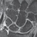
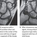
Quantitative CT (QCT) offers the advantage of selective determination of the density and thickness of both cortical and cancellous bone ( Fig. 31.4 ). In contrast to the previously described planar procedures, QCT computes the cancellous and cortical density as a real, volume-related unit from the directly calculated linear attenuation coefficient as a hydroxyapatite equivalent (mg/cm3). Osteodensitometry with peripheral QCT (pQCT) is very precise. The method measures additional parameters such as the thickness of the cortical wall and bone stability. This method is usually performed as single-energy QCT (SEQCT) at 80kV. The low voltage reduces error caused by overlying soft tissue. Here, the two-energy technique (DEQCT = dual-energy QCT) has only limited availability. The radiation exposure is considerably higher, and the reliability is lower.
Peripheral quantitative CT (pQCT) measures the bone mineral density of the distal radius or proximal tibia. The results are calculated in g/cm3. The effective equivalent dose is 1 μSv. The average statistical values of several German centers are listed in Table 31.4 .

The calculated bone density is not correlated to the age, but is recorded as a T score ( Fig. 31.5 ). The bone density is considered to be reduced if the mineral density is between 1 and 2.5 standard deviations (SD) below the average maximal value of a young, healthy adult of the same sex. Osteoporosis is diagnosed if the SD is–2.5 or below. This procedure takes into account that an agerelated T score does not represent the real risk. About 30% of postmenopausal women have an increased risk of fracture although their bone mineral density is normal for their age.

Quantitative ultrasonography examination (QUS) is performed on peripheral parts of the skeleton (phalanges, patella, tibia, calcaneus). This is not a measurement of bone density per se. Sound attenuation and changes in the sound velocity are defined as a function of changed bone mass or structure. A direct comparison with absorptiometric results is not possible. Here only indicative, indirect correlations are possible, such as the incidence of osteoporotic fractures in the subjects examined. The clinical validity has not been confirmed. At present, the procedure cannot be recommended for general use, although the approach is promising. It offers the advantages that it is uncomplicated, the equipment is portable, the costs are low, and no ionizing radiation is involved.
Quantitative MRI (QMR) determines the cancellous bone microstructure. This requires high-resolution MR images. The cancellous trabeculae cannot be directly visualized because of their lack of fat or water. The different magnetic susceptibility of osseous structures in the bone marrow causes inhomogeneities in the local magnetic field. The T2* relaxation time is inversely proportional to the density of the osseous structure, regardless of the bone mineral-salt content. The bone morphology determined is an important prognostic factor for the risk of fracture. The method is only possible on the peripheral skeleton (radius, fingers, calcaneus) because of the high spatial resolution required. Because of the sophisticated equipment and technical evaluation required, the procedure is at present performed only for research purposes.
Comparison of the measurement methods described is limited, for absorptiometric and radiographicmethods as well as those in nuclear medicine. This is especially true for procedures using ionizing radiation, as opposed to ultrasonography or MRI examinations. Further problems arise from the different results achieved among instruments produced by different manufacturers. In addition, bone density in a single region is not necessarily representative of the condition of the rest of the skeleton.
The different procedures are only indirectly appropriate for the determination of an absolute value. What is important is how bone density progresses with therapy which, considering the limitations of the individual procedures discussed above, should always be evaluated with the same instrument on the same part of the skeleton. Preference should be given pDXA for investigations of the distal radius. Clinical experience with this procedure is extensive, and the radiation exposure in the periphery is very lowwith an effective equivalent dose of 1 μSv.
A current approach is based on the relationship between reduced muscle strength and osteoporosis. The grip strength, measured objectively with a dynamometer, correlates significantly with bone mineral density. This simple method may allow to identify at-risk patients, who should then undergo further diagnostic procedures.
Therapeutic Options
The appropriate therapy is based on the endocrine, metabolic, or nutritive underlying disease. A calcium-rich diet, mobilization, physiotherapy, and analgesics are forms of symptomatic therapy. Medical therapy of osteoporosis includes:
1000–1500mg calcium daily
1000 IU vitamin D3 daily
Calcitonin (which inhibits the osteoclasts and relieves osteoporotic pain)
Fluoride (which stimulates the osteoblasts)
Bisphosphonates (in cases of rapid resorption of bone)
Estrogens (which have an antiresorptive effect; usually given together with a gestagen to counteract possible induction of endometrial carcinoma)
Therapeutic success is monitored by osteodensitometry.
Related posts:
Stay updated, free articles. Join our Telegram channel

Full access? Get Clinical Tree


