Case 32
Indication: Screening.
History: Bilateral implants 13 years previously.
Risk profile: No increased risk.
Age: 53 years.
Clinical Findings
Clinical inspection unremarkable. Implants appear “close-fitting”; within the tissue bilaterally. No pathological findings on palpation.
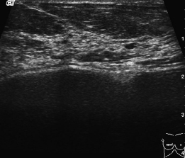
Fig. 32.1 Sonography, right breast.
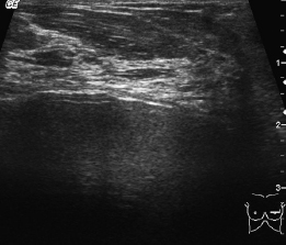
Fig. 32.2 Sonography, left breast.
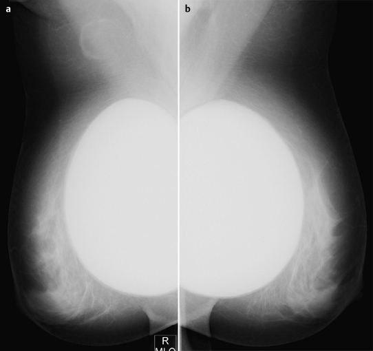
Fig. 32.3a,b Digital mammography, MLO view.
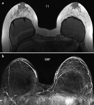
Fig. 32.4a,b Contrast-enhanced MR mammography.
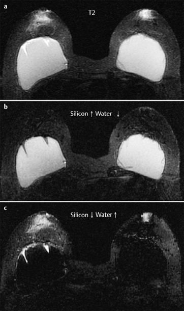
Fig. 32.5a–c MR mammography-prosthesis protocol.

|
Please characterize ultrasound, mammography, and MRI findings.
What is your preliminary diagnosis?
What are your next steps? |


