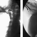
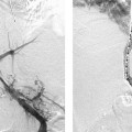
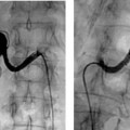
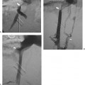
CASE 32 A 48-year-old male with end-stage renal disease presented to interventional radiology for removal of a clot from his left upper extremity arteriovenous graft. Figure 32-1 Treatment of rupture of the venous outflow of a left arm arterovenous graft. (A) Venography after anticoagulation, thrombolysis, and venoplasty of the venous outflow shows marked active extravasation. (B) Repeat venography after placement of a covered Viabahn stent (W. L. Gore, Newark, Delaware) shows resolution of the extravasation and no residual venous outflow stenosis. Flow within the graft was restored after thrombolysis with 4 mg recombinant tissue plasminogen activator, anticoagulation with 5000 IU heparin, and clearance of the graft using a 6-mm balloon across the arterial limb and a 7-mm balloon across the venous limb. A tight stenosis impeding clearance of the venous outflow was encountered, and the balloon dilated during the procedure. Venography performed immediately after balloon dilatation showed marked contrast extravasation at the site of venoplasty. Prolonged balloon tamponade failed to arrest hemorrhage. Rupture of the venous outflow of a dialysis shunt after venoplasty. The balloon catheter was exchanged over a standard guidewire for an 8-French (F) vascular sheath. A 5-cm long, 8-mm diameter covered stent (Viabahn, W. L. Gore, Newark, Delaware) was placed across the site of extravasation. Following deployment, the stent was dilated with an 8-mm balloon. Follow-up venography showed no residual extravasation and no residual stenosis within the venous outflow. 8F vascular sheath 8-mm diameter balloon catheter 5-cm long, 8-mm diameter Viabahn stent (W. L. Gore, Newark, Delaware) Contrast material
Clinical Presentation

Radiologic Studies
Diagnosis
Treatment
Equipment
Discussion
Background
Stay updated, free articles. Join our Telegram channel

Full access? Get Clinical Tree


