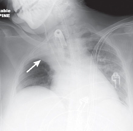
CASE 33 25-year-old woman, status post conversion of pre-existing endotracheal tube to a tracheostomy device, with an abrupt decrease in oxygen saturations and increased work of breathing immediately following the procedure AP chest radiograph (Fig. 33.1) demonstrates opacification of the upper one-third of the right hemithorax without air bronchograms. The horizontal fissure (arrow) is displaced cephalad. Various life support tubes and lines are appropriately positioned. Free air beneath the left diaphragm is related to the concurrent recent PEG placement. Right Upper Lobe Atelectasis (Resorption); Endobronchial Mucus Plug • Acute Lobar Collapse of Other Etiologies Fig. 33.1 Resorption atelectasis is the most common and complex mechanism of volume loss and results from obstruction in airflow somewhere between the trachea and alveoli. In acute endobronchial obstruction, the partial pressure of gases in mixed venous blood (Pp mixed venous blood) becomes less than that in alveolar air (Pp alveolar air). As blood passes through alveolar capillaries, the partial pressures of gases (Pp gases) equilibrate and oxygen (O2) diffuses into the capillary bed. Alveoli diminish in volume in proportion to the quantity of O2 absorbed. The partial pressures of alveolar carbon dioxide (Pp CO2) and nitrogen (Pp N2) then increase relative to those in the capillary bed. These gases also diffuse into the capillary bed to maintain gaseous equilibrium, with a proportional decrease in alveolar volume. The partial pressure of alveolar oxygen (Pp alveolar O2) then increases relative to the capillary bed. Oxygen again diffuses into the capillary bed to maintain equilibrium, with a resultant proportional decrease in alveolar volume. This cycle repeats over and over until eventually all alveolar gas is absorbed and the alveoli are completely collapsed (Fig. 33.2).
 Clinical Presentation
 Radiologic Findings
 Diagnosis
 Differential Diagnosis
 Aspirated or Dislodged Foreign Body (e.g., tooth, dental amalgam)
 Aspirated Peri-operative Blood/Endobronchial Clot

 Discussion
Background
Clinical Findings
Stay updated, free articles. Join our Telegram channel

Full access? Get Clinical Tree


