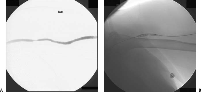
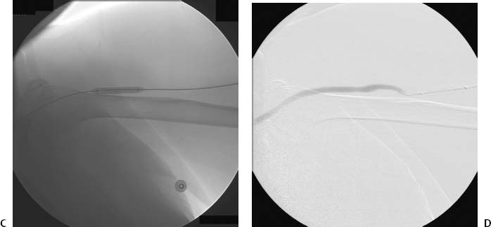
CASE 34 A 43-year-old female with an upper arm dialysis fistula was referred to interventional radiology for evaluation of high venous pressures at dialysis. Physical examination of the arm revealed a patent fistula with a bounding pulse. Figure 34-1 Prophylactic angioplasty of venous stenosis in dialysis fistula. (A) Initial digital subtraction venogram shows venous stenosis at the level of the mid humerous. (B) Fluoroscopic image shows high-pressure angioplasty balloon being inflated across the lesion. (C) Fluoroscopic image shows high-pressure angioplasty balloon completely inflated across the lesion. (D) Follow-up digital subtraction venogram after balloon angioplasty shows wide patency of venous outflow. The fistula was punctured using a 21-gauge needle from a Micropuncture set (Cook, Bloomington, Indiana), and the needle was exchanged over a 0.018″ guidewire for a 5-French (F) dilator that was used to perform fistulography. The fistulagram revealed a high-grade stenosis of the outflow vein at the level of the upper humerus (Fig. 34-1A). Venous outflow stenosis. A 7-mm × 4-cm high-pressure angioplasty balloon (Blue Max, Boston Scientific, Natick, Massachusetts) was advanced to the stenosis and inflated to 16 atmospheres (Fig. 34-1B). The balloon was completely inflated without a waist to indicate residual stenosis (Fig. 34-1C). Follow-up venogram showed wide patency of the segment without residual stenosis (Fig. 34-1D). High-pressure angioplasty balloon catheters Micropuncture set (Cook, Bloomington, Indiana) Soft-tipped 0.035″ guidewires Contrast material
Clinical Presentation
Radiologic Studies
Diagnosis
Treatment
Equipment
Discussion
Background
Related posts:


Stay updated, free articles. Join our Telegram channel

Full access? Get Clinical Tree


