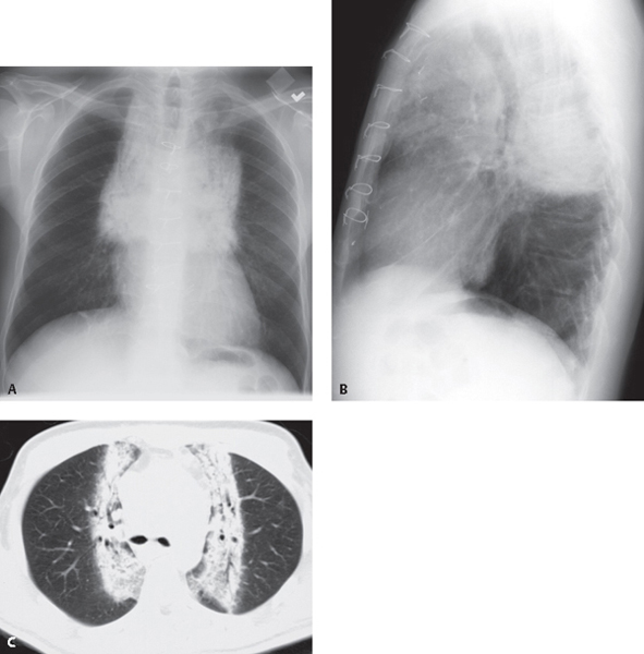
CASE 35 51-year-old man who is now five months status post mantle radiation therapy for unresectable adenocarcinoma of the lung PA (Fig. 35.1A) and lateral (Fig. 35.1B) chest radiographs demonstrate well-defined opacities with sharp borders involving the superior mediastinum and paramediastinal lung zones. The bronchi within the affected lung are distorted and dilated. Note the hilar retraction, diaphragmatic elevation, and compensatory overinflation of the uninvolved lower lungs. Chest CT (lung window) (Fig. 35.1C) reveals sharply demarcated bands of increased attenuation in the medial anterior and posterior paramediastinal lung zones. Note the traction bronchiectasis and bronchovascular reorientation in the affected lung. Fig. 35.1 Cicatrization Atelectasis: Iatrogenic Radiation-Therapy Induced; Corresponding to the Radiation Therapy Portal None Localized cicatrization atelectasis
 Clinical Presentation
 Radiologic Findings
 Diagnosis
 Differential Diagnosis
 Discussion
Background
![]()
Stay updated, free articles. Join our Telegram channel


Full access? Get Clinical Tree


