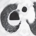
CASE 36 1,260 g girl born at 30 weeks’ gestation presenting with retractions, grunting, and poor air movement and requiring oxygen; has received one dose of surfactant, with some clinical improvement AP chest radiograph (Fig. 36.1) shows bilateral, symmetrical, perihilar opacities with a fine granular pattern and associated air bronchograms. The lung volumes are diminished bilaterally. Note the endotracheal tube, enteric tube, umbilical arterial and umbilical venous catheters. Adhesive Atelectasis: Respiratory Distress Syndrome (RDS) of the Newborn • Pneumonia • Volume overload or heart failure • Massive aspiration • Pulmonary hemorrhage The pressure-volume relationships of the lung depend on forces acting at the air-tissue interface of the alveolar wall. These forces are further understood by applying Laplace’s law (p = 2T/r), where p represents pressure, T the surface tension at the air-tissue interface, and r
 Clinical Presentation
 Radiologic Findings
 Diagnosis
 Differential Diagnosis
 Discussion
Background
![]()
Stay updated, free articles. Join our Telegram channel

Full access? Get Clinical Tree


