
MRM score | Finding right | Points |
Shape | round | 0 |
Border | well-defined | 0 |
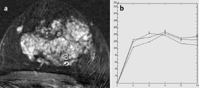
CM Distribution | inhomogenous | 1 |
Initial Signal Intensity Increase | strong | 2 |
Post-initial Signal Intensity Character | wash-out | 2 |
MRI score (points) |
| 5 |
MRI BI-RADS |
| 4 |
 Preliminary Diagnosis
Extensive adenosis of the left breast.
Differential Diagnosis
Diffuse breast cancer.
Clinical Findings | right- | left 1 |
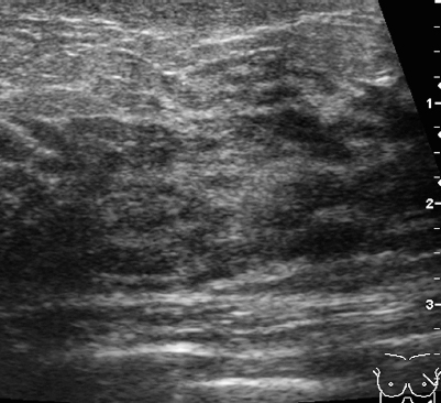
Ultrasound | right- | left 1 |
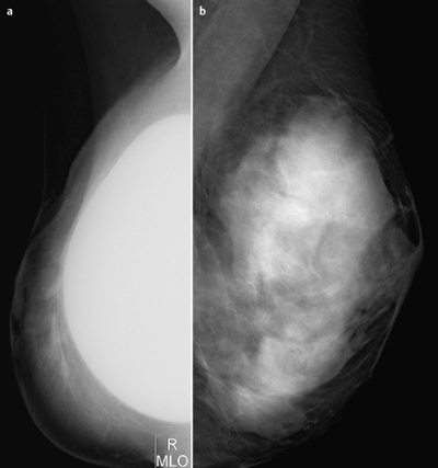
Mammography | right- | left 1 |
MR Mammography | right- | left 4 |
BI-RADS Total | right- | left 4 |
Procedure
Follow-up MRI in a more suitable week of the menstrual cycle.
Next Step
US-guided blind biopsy (14 gauge) of the left breast at 1 -o’clock,3-o’clock and 5-o’clock positions.
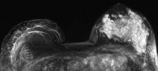
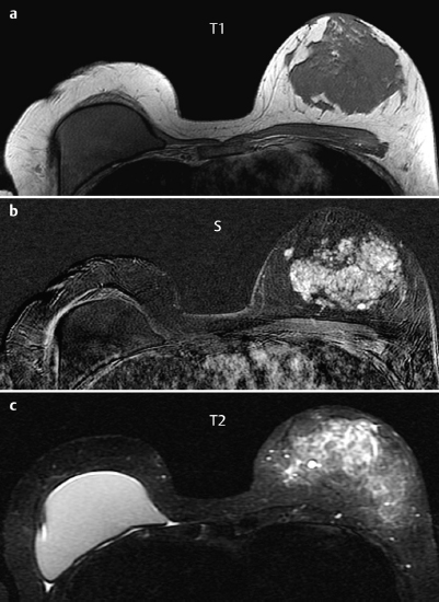
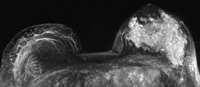
Fig. 37.6 Original MR mammography. Menstrual cycle week 4.
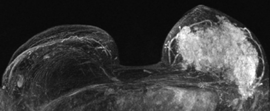
Fig. 37.7 Follow-up MR mammography. Menstrual cycle week 2. Enhancement pattern identical to primary MRI.
Histology of the left breast (3 biopsy specimens)
Adenosis.

Stay updated, free articles. Join our Telegram channel

Full access? Get Clinical Tree


