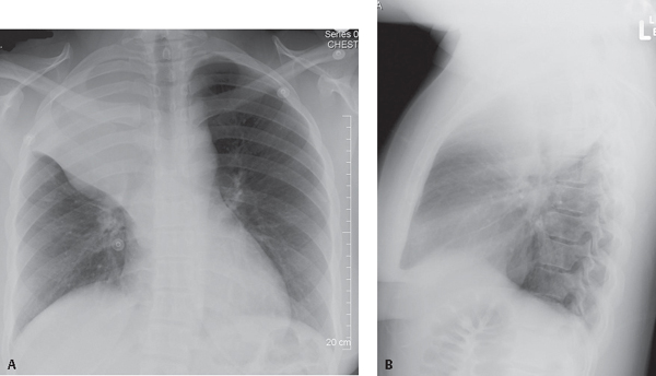
CASE 38 27-year-old woman complains of non-productive cough and weight loss over the last several months PA (Fig. 38.1A) and lateral (Fig. 38.1B) chest X-rays reveal reduction in volume of the right hemithorax, right hemidiaphragm elevation, and a “peaked” appearance (i.e., juxtaphrenic peak) of the superomedial hemidiaphragm. The trachea is minimally displaced to the right. On the frontal exam (Fig. 38.1A), the horizontal fissure is displaced cephalad. A mass-like convex-appearing bulge is present in the inferior and medial portion of the fissure. The lateral aspect of the fissure is concave superiorly. The configuration creates a “reverse S-shaped” morphology to the fissure. The lateral exam (Fig. 38.1B) reveals displacement of both the horizontal and oblique fissures and an indistinct wedge-shaped triangular mediastinal opacity with its apex directed toward the hilum and its base contiguous with the parietal pleura posterior to the apex of the hemithorax. Complicated Right Upper Lobe Atelectasis; Lymphoma with “Reverse ‘S’ Sign of Golden” Fig. 38.1 Fig. 38.2. (A) PA and (B)
 Clinical Presentation
 Radiologic Findings
 Diagnosis
Related posts:
![]()
Stay updated, free articles. Join our Telegram channel

Full access? Get Clinical Tree


38 Complicated Right Upper Lobe Atelectasis: Reverse “S” Sign of Golden


