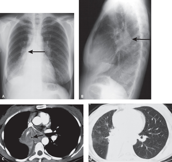
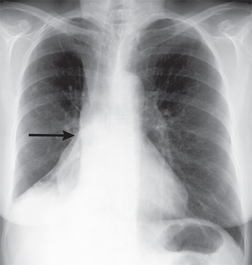
CASE 39 37-year-old woman with new onset of seizures and brain metastases PA (Fig. 39.1A) and lateral (Fig. 39.1B) chest radiographs show a convex perihilar mass (arrow) medial to the bronchus intermedius obliterating the right lower lobe bronchus origin. There is inferior displacement of the right hilum and oblique fissure. The right interlobar artery and proximal lower lobe bronchus are inconspicuous. A positive spine sign is present on the lateral exam manifest by increased opacity over the lower thoracic spine which silhouettes the posterior right diaphragm. Note the right diaphragm elevation on both frontal and lateral chest radiography. Contrast-enhanced chest CT (Fig. 39.1C, mediastinal window, and Fig. 39.1D, lung window) demonstrates a heterogeneous subcarinal and right infrahilar mass encasing the bronchus intermedius (arrow) and interlobar artery. The middle lobe bronchi are narrowed but patent. There is obliteration of the lower lobe bronchi with distal atelectasis and consolidation as well as inferior, medial, and posterior displacement of the oblique fissure. Fig. 39.1 Right Lower Lobe Atelectasis; Obstructing Small Cell Lung Cancer • Post-Obstructive Lower Lobe Atelectasis from an Endobronchial Mass (e.g., primary and secondary neoplasia) • Post-Obstructive Lower Lobe Atelectasis from Extrinsic Bronchial Compression by Reactive or Neoplastic Lymphadenopathy The right and left lower lobe demonstrate similar patterns of lobar collapse on imaging. The triangular versus rounded configuration assumed by the atelectatic lower lobe is related to the fulcrum-like effect exerted on the lung by the hilum and the integrity of the pulmonary ligament. When the pulmonary ligament is complete, the lower lobe is tethered to the mediastinum and hemidiaphragm and maintains a close relationship to both structures, assuming a triangular configuration as it loses volume (Fig. 39.2). Alternatively, when the pulmonary ligament is incomplete, the lung base is not adherent to the ipsilateral hemidiaphragm. As the lower lobe looses volume, the resultant shape depends primarily on its mediastinal attachment and the collapsed lobe assumes a more rounded morphology (Fig. 39.3). Fig. 39.2
 Clinical Presentation
 Radiologic Findings
 Diagnosis
 Differential Diagnosis
 Discussion
Background
Related posts:
![]()
Stay updated, free articles. Join our Telegram channel

Full access? Get Clinical Tree


Radiology Key
Fastest Radiology Insight Engine


