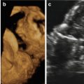
Fig. 17.1
Three-dimensional ultrasound in surface-rendering mode showing proboscis and severe hypotelorism
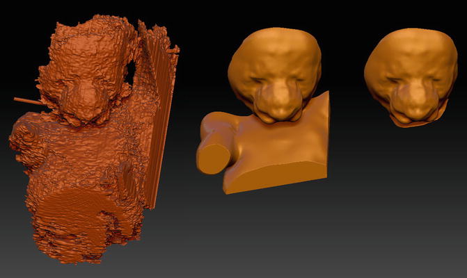
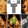
Fig. 17.2
3D virtual physical model showing the typical features of the congenital malformation
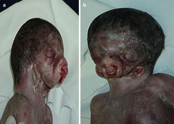
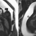
Fig. 17.3
Postmortem photographs showing the agnathia/otocephaly complex in lateral (a) and frontal (b) views confirming the accuracy of the prenatal ultrasound diagnosis
This work was based on a previously published report [4].
References
1.
2.
3.
4.
Menezes GA, Araujo Junior E, Lopes J, Belmonte S, Tonni G, Werner H. Prenatal diagnosis and physical model reconstruction of agnathia-otocephaly with limb deformities (absent ulna, fibula and digits) following maternal exposure to oxymetazoline in the first trimester. J Obstet Gynecol Research. 2016;42:1016–20.
Related posts:
 Congenital Subcutaneous Mixed Venous-Lymphatic Orofacial Malformation Associated with Macroglossia: Prenatal Diagnosis with Ultrasound and Fetal MRI
 The Genetics of Facial Cleft
 Acromelic Frontonasal Dysplasia (Median Cleft Face Syndrome)
 The Fetal Brain in Fetuses with Orofacial Abnormalities
 Magnetic Resonance Imaging (MRI) in the Evaluation of the Fetal Face
 The Role of 2D/3D/4D Ultrasound in the Prenatal Assessment of Cleft Lip and Palate
Stay updated, free articles. Join our Telegram channel

Full access? Get Clinical Tree


