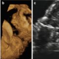
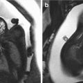
Fig. 15.1
Three-dimensional ultrasound at 16 weeks 5 days of gestation in the axial plane (a) and surface rendering mode (b) demonstrating an exophytic midline lesion originating from the brain and protruding through the nasal cavity

Fig. 15.2
Three-dimensional (3D) ultrasound in the coronal plane using “skeleton” mode: a midline fronto-ethmoid bony defect was clearly detected and rendered

Fig. 15.3
Three-dimensional ultrasound using tomographic ultrasound imaging (TUI) in the coronal plane with slice thickness of 1 mm: a hyperechogenic tumor with undefined contour is seen projecting anteriorly through the nasal cavity. Severe hypertelorism was an associated finding

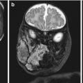
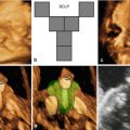
Fig. 15.4
(a, b). Fetal T2-weighted MRI at 19 weeks 5 days of gestation showed the homogeneous hyperintense lesion with corresponding heterogeneous hyperintense signal on T2-weighted images

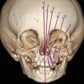
Fig. 15.5
(a) Postmortem CT scan with (b) 3D rendering confirming the prenatal US findings of fronto-ethmoid bony defect

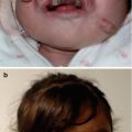
Fig. 15.6
(a) Postmortem CT scan with 3D rendering and (b) gross pathology

Fig. 15.7
 Congenital Subcutaneous Mixed Venous-Lymphatic Orofacial Malformation Associated with Macroglossia: Prenatal Diagnosis with Ultrasound and Fetal MRI
 The Genetics of Facial Cleft
 Median Cleft Lip and Palate, Cutaneous Nasal Polyps, and Corpus Callosum Lipoma: A Case of Pai Syndrome Associated with Ventricular Septal Defects
 The Fetal Brain in Fetuses with Orofacial Abnormalities
 Magnetic Resonance Imaging (MRI) in the Evaluation of the Fetal Face
 The Role of 2D/3D/4D Ultrasound in the Prenatal Assessment of Cleft Lip and Palate


Neuropathology confirmed the presence of neural tissue within the tumor (arrow)
Related posts:
Congenital Subcutaneous Mixed Venous-Lymphatic Orofacial Malformation Associated with Macroglossia: Prenatal Diagnosis with Ultrasound and Fetal MRI
The Genetics of Facial Cleft
Median Cleft Lip and Palate, Cutaneous Nasal Polyps, and Corpus Callosum Lipoma: A Case of Pai Syndrome Associated with Ventricular Septal Defects
The Fetal Brain in Fetuses with Orofacial Abnormalities
Magnetic Resonance Imaging (MRI) in the Evaluation of the Fetal Face
The Role of 2D/3D/4D Ultrasound in the Prenatal Assessment of Cleft Lip and Palate
Stay updated, free articles. Join our Telegram channel
Full access? Get Clinical Tree
