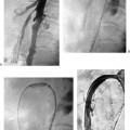
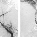
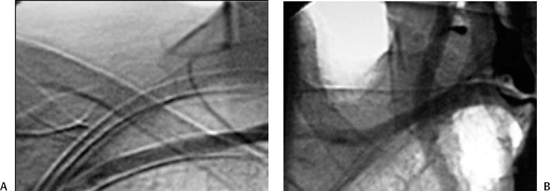
CASE 40 A 45-year-old female presented with intermittent dysphasia to solids for the past year and a 20-pound weight loss. Physical exam and laboratory tests were otherwise normal. Figure 40-1 Combined esophagogram and aortogram in a 45-year-old female with dysphagia. (A) In the frontal projection, the right subclavian artery appears to originate from the aortic arch to the left of the right common carotid artery. (B) Aortogram in the lateral projection after oral administration of a capful of barium contrast material shows characteristic compression of the posterior aspect of the esophagus by the aberrant right subclavian (lusoria) artery. Chest radiography was unremarkable. After swallowing a capful of high-density barium, the patient underwent aortography. The study revealed a left aortic arch with a replaced right subclavian artery arising distal to the left subclavian artery. This vessel passed behind and partially compressed the esophagus. Dysphagia lusoria resulting from a replaced right subclavian artery. The patient underwent surgical correction from the supraclavicular approach. The right subclavian artery origin was ligated, and the artery was transected. An end-to-side anastomosis was then created between the right subclavian artery and the right common carotid artery. After 1 year, the patient remains symptom-free. Dysphagia lusoria, or “dysphagia by freak of nature,” is an unfortunate term used to describe difficulty swallowing caused by compression of the esophagus by a vascular ring. The aberrant right subclavian (“lusoria”) artery (ARSA) arises from a normally positioned left aortic arch in approximately 1 in 200 individuals and represents the most common congenital aortic arch abnormality. The embryologic origin involves involution of the fourth vascular arch and the right dorsal aorta, leaving the seventh intersegmental artery arising from the descending aorta to supply the arm (the ARSA). In the vast majority of cases, the ARSA is retroesophageal, but in rare cases, it may pass anterior to the esophagus. ARSA does not result in a complete vascular “ring” because the compressed structures are not completely encircled by anomalous vasculature. An aneurysm of the ARSA and its aortic origin may increase the likelihood of compressive symptoms and has been termed the “diverticulum of Kommerell.”
Clinical Presentation
Radiologic Studies
Diagnosis
Treatment
Discussion
Background
Stay updated, free articles. Join our Telegram channel

Full access? Get Clinical Tree


