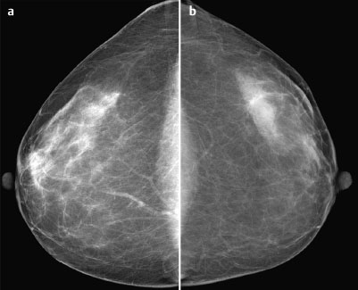
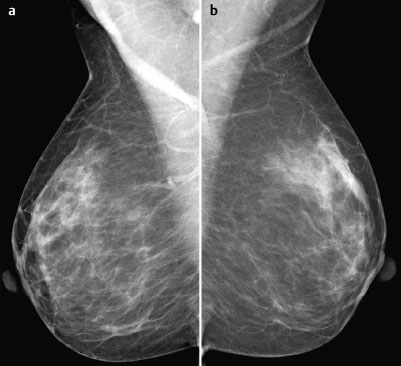
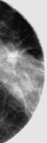
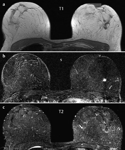
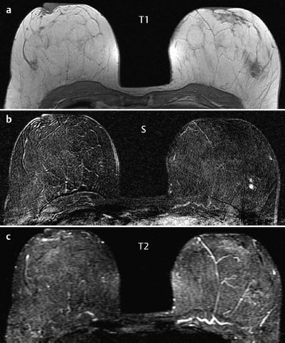
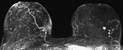
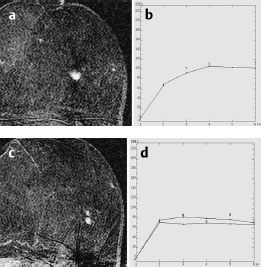
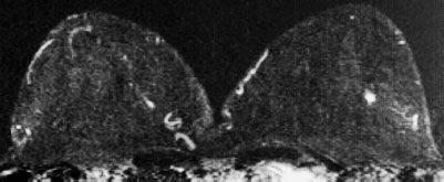
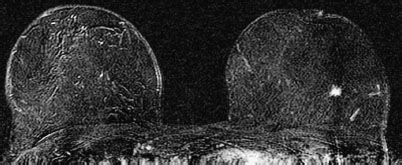
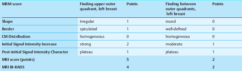
Case 42 Indication: Screening mammography. History: Unremarkable. Risk profile: Normal. Age: 54 years. No findings. Fig. 42.1 Ultrasound. Fig. 42.2a,b Digital mammography, CC view. Fig.42.3a,b Digital mammography, MLO view. Fig. 42.4 Spot compression, left breast. Fig. 42.5a-c Contrast-enhanced MRI of the breasts. Fig. 42.6a-c Contrast-enhanced MRI of the breasts. Fig. 42.7 Contrast-enhanced MR mammography. Maximum intensity projection. Fig. 42.8a-d Signal-to-time curves. Please characterize ultrasound, mammography, and MRI findings. What is your preliminary diagnosis? What are your next steps? This case demonstrates the imaging studies of an asymptomatic woman presenting for screening. In the upper outer quadrant of the left breast there was an irregular, hypoechoic lesion with distal acoustic shadowing (diameter8 mm). US BI-RADS left 4. MRI demonstrated a spiculated, ill-defined mass (diameter: 1 cm) in the upper outer quadrant with strong initial signal increase and postinitial plateau as well as a reduced signal in T2-weighted imaging. 1.2 cm caudal of this mass there were two homogeneously enhancing lesions, each of 5 mm diameter, with initial signal increase of 80% and postinitial plateau as well as reduced signal in T2-weighted imaging (MRI BI-RADS 3). Imaging showed an asymmetric (right>left), partially inhomogeneous dense parenchyma, ACR type 3. There was an isodense, slightly spiculated mass (diameter 1 cm) in the upper outer quadrant of the left breast. Spot compression obtained a more precise depiction of the spiculated quality of this lesion. BI-RADS right 1/left 4. PGMI: CC view P; MLO view G (inframammary fold). The lesion in the upper outer quadrant of the left breast was visible, at a similar size, in MR images produced 3 years previously in another clinic (Fig. 42.9). MRI Artifact Category: 2 MRI Density Type: 1 Fig. 42.9 MR mammography three years earlier demonstrated a hypervascularized lesion in the left breast [imaging not performed by authors]. 256 matrix. Fig. 42.10 Current MR mammography for comparison. 512 matrix. Left upper outer quadrant: Carcinoma. Left between outer quadrants: Fibroadenoma. Adenosis, papilloma.
Clinical Findings
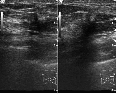

Ultrasound
MR Mammography
Mammography
Note
 Preliminary Diagnosis
 Differential Diagnosis
BI-RADS Categorization | ||
Clinical Findings | right 1 | left 1 |
Ultrasound | right 1 | left 4 |
Mammography | right 1 | left 4 |
MR Mammography | right 1 | left 4 |
BI-RADS Total | right 1 | left 4 |
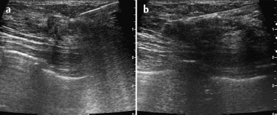
Fig. 42.11 a,b US-guided core biopsy of the left breast. Pre-fire and post-fire documentation.
Procedure
US-guided core biopsy and histopathological evaluation of the lesion in the upper outer quadrant of the left breast (Fig. 42.11a,b).
Histopathology of the core biopsy specimen
Tubular carcinoma.
Further procedure
Open biopsy of the tubular carcinoma as well as of the secondary lesions in the left breast after MRI-guided hook-wire localization(Fig. 42.12).
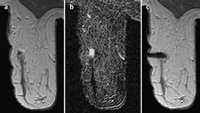
Fig. 42.12a-c MR-guided preoperative hook-wire localization. Pre-contrast image, subtraction image and documentation of the wire inposition.
Histology
Tubular carcinoma (diameter 12 mm) accompanied by focal adenosis.
TC pTIc, pN1,G1.
Stay updated, free articles. Join our Telegram channel

Full access? Get Clinical Tree


